 Movie
Movie Controller

[English] 日本語
 Yorodumi
- PDB-1e19: Structure of the carbamate kinase-like carbamoyl phosphate synthe... -
+ Open data

- Basic information
| Entry | Database: PDB / ID: 1.0E+19 | ||||||
|---|---|---|---|---|---|---|---|
| Title | Structure of the carbamate kinase-like carbamoyl phosphate synthetase from the hyperthermophilic archaeon Pyrococcus furiosus bound to ADP | ||||||
 Components Components | CARBAMATE KINASE | ||||||
 Keywords Keywords | TRANSFERASE / HYPERTHERMOPHILES / ADP SITE / ARGININE METABOLISM PHOSPHORYL GROUP TRANSFER | ||||||
| Function / homology |  Function and homology information Function and homology informationcarbamate kinase / carbamate kinase activity / : / ATP binding / cytosol Similarity search - Function | ||||||
| Biological species |   PYROCOCCUS FURIOSUS (archaea) PYROCOCCUS FURIOSUS (archaea) | ||||||
| Method |  X-RAY DIFFRACTION / SYNCHROTRON / MOLECULAR REPLACEMENT / Resolution: 1.5 Å X-RAY DIFFRACTION / SYNCHROTRON / MOLECULAR REPLACEMENT / Resolution: 1.5 Å | ||||||
 Authors Authors | Ramon-Maiques, S. / Marina, A. / Uriarte, M. / Fita, I. / Rubio, V. | ||||||
 Citation Citation | Journal: J.Mol.Biol. / Year: 2000 Title: The 1.5-A Resolution Crystal Structure of the Carbamate Kinase-Like Carbamoyl Phosphate Synthetase from the Hyperthermophilic Archaeon Pyrococcus Furiosus, Bound to Adp, Confirms that This ...Title: The 1.5-A Resolution Crystal Structure of the Carbamate Kinase-Like Carbamoyl Phosphate Synthetase from the Hyperthermophilic Archaeon Pyrococcus Furiosus, Bound to Adp, Confirms that This Thermoestable Enzyme is a Carbamate Kinase, and Provides Insights Into Substrate Binding and Stability in Carbamate Kinases Authors: Ramon-Maiques, S. / Marina, A. / Uriarte, M. / Fita, I. / Rubio, V. | ||||||
| History |
|
- Structure visualization
| Structure viewer | Molecule: MolmilJmol/JSmol |
|---|
- Downloads & links
-Download
| PDBx/mmCIF format | 1e19.cif.gz | 154.7 KB | Display | PDBx/mmCIF format |
|---|---|---|---|---|
| PDB format | pdb1e19.ent.gz | 119.9 KB | Display | PDB format |
| PDBx/mmJSON format | 1e19.json.gz | Tree view | PDBx/mmJSON format | |
| Others |  Other downloads Other downloads |
-Validation report
| Arichive directory | https://data.pdbj.org/pub/pdb/validation_reports/e1/1e19ftp://data.pdbj.org/pub/pdb/validation_reports/e1/1e19 | HTTPS FTP |
|---|
-Related structure data
| Related structure data | 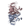 1b7bS S: Starting model for refinement |
|---|---|
| Similar structure data |
-Links
 PDBj
PDBj- Assembly
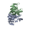
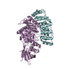
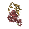
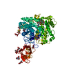
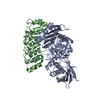
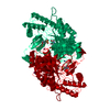
| Deposited unit | 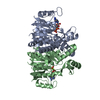
| ||||||||
|---|---|---|---|---|---|---|---|---|---|
| 1 |
| ||||||||
| Unit cell |
| ||||||||
| Noncrystallographic symmetry (NCS) | NCS oper: (Code: given Matrix: (0.958996, -0.047676, 0.279105), Vector: |
-Components
| #1: Protein | Mass: 34475.582 Da / Num. of mol.: 2 / Source method: isolated from a natural source / Source: (natural) PYROCOCCUS FURIOSUS (archaea) / References: UniProt: P95474, carbamate kinase#2: Chemical |   Mass: 427.201 Da / Num. of mol.: 2 / Source method: obtained synthetically / Formula: C10H15N5O10P2 / Comment: ADP, energy-carrying molecule*YM Mass: 427.201 Da / Num. of mol.: 2 / Source method: obtained synthetically / Formula: C10H15N5O10P2 / Comment: ADP, energy-carrying molecule*YM#3: Chemical |   Mass: 24.305 Da / Num. of mol.: 2 / Source method: obtained synthetically / Formula: Mg Mass: 24.305 Da / Num. of mol.: 2 / Source method: obtained synthetically / Formula: Mg#4: Water | ChemComp-HOH / |  Mass: 18.015 Da / Num. of mol.: 716 / Source method: isolated from a natural source / Formula: H2O Mass: 18.015 Da / Num. of mol.: 716 / Source method: isolated from a natural source / Formula: H2O |
|---|
-Experimental details
-Experiment
| Experiment | Method: X-RAY DIFFRACTION / Number of used crystals: 1 |
|---|
- Sample preparation
| Crystal | Density Matthews: 2.5 Å3/Da / Density % sol: 50 % | ||||||||||||||||||||||||||||||||||||||||||
|---|---|---|---|---|---|---|---|---|---|---|---|---|---|---|---|---|---|---|---|---|---|---|---|---|---|---|---|---|---|---|---|---|---|---|---|---|---|---|---|---|---|---|---|
| Crystal grow | pH: 8 Details: 1.3M SODIUM CITRATE 0.1M TRIS-HCL, PH=8.5, 0-10% ETHYLENE GLYCOL PROTEIN SOLUTION: 10MG/ML OF PROTEIN IN 10MM TRIS-HCL CONTAINING 20MM ATP AND MGCL2, pH 8.00 | ||||||||||||||||||||||||||||||||||||||||||
| Crystal grow | *PLUS Temperature: 22 ℃ / Method: vapor diffusion, hanging drop / pH: 8.5 | ||||||||||||||||||||||||||||||||||||||||||
| Components of the solutions | *PLUS
|
-Data collection
| Diffraction | Mean temperature: 100 K |
|---|---|
| Diffraction source | Source: SYNCHROTRON / Site: ESRF  / Type: ESRF / Wavelength: 0.93 / Type: ESRF / Wavelength: 0.93 |
| Detector | Type: MARRESEARCH / Detector: CCD / Date: Sep 15, 1998 |
| Radiation | Protocol: SINGLE WAVELENGTH / Monochromatic (M) / Laue (L): M / Scattering type: x-ray |
| Radiation wavelength | Wavelength: 0.93 Å / Relative weight: 1 |
| Reflection | Resolution: 1.43→76.67 Å / Num. obs: 116569 / % possible obs: 97.3 % / Redundancy: 3.8 % / Rmerge(I) obs: 0.067 / Rsym value: 0.067 / Net I/σ(I): 5.2 |
| Reflection shell | Resolution: 1.5→1.59 Å / Rmerge(I) obs: 0.355 / Mean I/σ(I) obs: 1.9 / Rsym value: 0.355 / % possible all: 98.6 |
| Reflection | *PLUS Num. measured all: 448640 |
| Reflection shell | *PLUS Highest resolution: 1.5 Å / % possible obs: 98.6 % |
- Processing
| Software |
| |||||||||||||||||||||||||||||||||||||||||||||||||||||||||||||||
|---|---|---|---|---|---|---|---|---|---|---|---|---|---|---|---|---|---|---|---|---|---|---|---|---|---|---|---|---|---|---|---|---|---|---|---|---|---|---|---|---|---|---|---|---|---|---|---|---|---|---|---|---|---|---|---|---|---|---|---|---|---|---|---|---|
| Refinement | Method to determine structure: MOLECULAR REPLACEMENT Starting model: PDB ENTRY 1B7B Resolution: 1.5→15 Å / Cross valid method: THROUGHOUT / σ(F): 0
| |||||||||||||||||||||||||||||||||||||||||||||||||||||||||||||||
| Refinement step | Cycle: LAST / Resolution: 1.5→15 Å
| |||||||||||||||||||||||||||||||||||||||||||||||||||||||||||||||
| Refine LS restraints |
| |||||||||||||||||||||||||||||||||||||||||||||||||||||||||||||||
| Software | *PLUS Name: REFMAC / Classification: refinement | |||||||||||||||||||||||||||||||||||||||||||||||||||||||||||||||
| Refinement | *PLUS Rfactor obs: 0.183 | |||||||||||||||||||||||||||||||||||||||||||||||||||||||||||||||
| Solvent computation | *PLUS | |||||||||||||||||||||||||||||||||||||||||||||||||||||||||||||||
| Displacement parameters | *PLUS | |||||||||||||||||||||||||||||||||||||||||||||||||||||||||||||||
| Refine LS restraints | *PLUS
| |||||||||||||||||||||||||||||||||||||||||||||||||||||||||||||||
| LS refinement shell | *PLUS Highest resolution: 1.5 Å / Lowest resolution: 1.66 Å / Rfactor Rfree: 0.267 / Num. reflection Rfree: 1593 / Num. reflection obs: 24677 / Rfactor obs: 0.229 |
