- PDB-2w9r: Structural basis of N-end rule substrate recognition in Escherich... -
+
Open data
ID or keywords:
Loading...
-
Basic information
Entry
Database: PDB / ID: 2w9r
Title
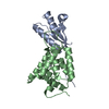
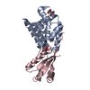
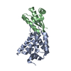
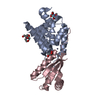
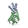
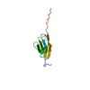
Structural basis of N-end rule substrate recognition in Escherichia coli by the ClpAP adaptor protein ClpS
Components
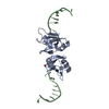
ATP-DEPENDENT CLP PROTEASE ADAPTER PROTEIN CLPS
DNA PROTECTION DURING STARVATION PROTEIN
Keywords
CHAPERONE / ADAPTOR PROTEIN / DNA CONDENSATION / IRON / CLPS / CLPA / CYTOPLASM / N-END RULE / DNA-BINDING / IRON STORAGE / METAL-BINDING / OXIDOREDUCTASE
Function / homology
Function and homology information
DnaA-Dps complex / Oxidoreductases; Oxidizing metal ions / oxidoreductase activity, acting on metal ions / nucleoid / chromosome condensation / molecular function inhibitor activity / response to starvation / response to stress / negative regulation of DNA-templated DNA replication initiation / ferric iron binding ...DnaA-Dps complex / Oxidoreductases; Oxidizing metal ions / oxidoreductase activity, acting on metal ions / nucleoid / chromosome condensation / molecular function inhibitor activity / response to starvation / response to stress / negative regulation of DNA-templated DNA replication initiation / ferric iron binding / protein catabolic process / protein-folding chaperone binding / response to heat / intracellular iron ion homeostasis / proteolysis / DNA binding / identical protein binding / membrane / cytoplasm Similarity search - Function
DNA protection during starvation protein, gammaproteobacteria / Ribosomal protein L7/L12, C-terminal domain/Adaptor protein ClpS / ATP-dependent Clp protease adaptor protein ClpS / Adaptor protein ClpS, core / ATP-dependent Clp protease adaptor protein ClpS / Dps protein family signature 2. / Dps protein family signature 1. / DNA-binding protein Dps, conserved site / DNA-binding protein Dps / Ribosomal Protein L30; Chain: A, ...DNA protection during starvation protein, gammaproteobacteria / Ribosomal protein L7/L12, C-terminal domain/Adaptor protein ClpS / ATP-dependent Clp protease adaptor protein ClpS / Adaptor protein ClpS, core / ATP-dependent Clp protease adaptor protein ClpS / Dps protein family signature 2. / Dps protein family signature 1. / DNA-binding protein Dps, conserved site / DNA-binding protein Dps / Ribosomal Protein L30; Chain: A, / Ribosomal protein L7/L12, C-terminal/adaptor protein ClpS-like / Ferritin/DPS protein domain / Ferritin-like domain / Ferritin-like / Ferritin-like superfamily / 2-Layer Sandwich / Alpha Beta Similarity search - Domain/homology
ATP-dependent Clp protease adapter protein ClpS / DNA protection during starvation protein Similarity search - Component
Biological species
ESCHERICHIA COLI (E. coli)
Method
X-RAY DIFFRACTION / SYNCHROTRON / OTHER / Resolution: 1.7 Å
Mass: 12321.168 Da / Num. of mol.: 1 Source method: isolated from a genetically manipulated source Details: CLPS PROTEIN FROM ESCHERICHIA COLI IN COMPLEX WITH LEU-PEPTIDE Source: (gene. exp.) ESCHERICHIA COLI (E. coli) / Strain: K12 / Production host: ESCHERICHIA COLI (E. coli) / Strain (production host): BL21 / References: UniProt: P0A8Q6
#2: Protein/peptide
DNAPROTECTIONDURINGSTARVATIONPROTEIN / PEXB / VTM
Mass: 1251.515 Da / Num. of mol.: 1 / Fragment: RESIDUES 10-16 Source method: isolated from a genetically manipulated source Source: (gene. exp.) ESCHERICHIA COLI (E. coli) / Strain: K12 / Production host: ESCHERICHIA COLI (E. coli) / Strain (production host): BL21 / References: UniProt: P0ABT2
Protocol: SINGLE WAVELENGTH / Monochromatic (M) / Laue (L): M / Scattering type: x-ray
Radiation wavelength
Wavelength: 0.97 Å / Relative weight: 1
Reflection
Resolution: 1.7→30 Å / Num. obs: 11540 / % possible obs: 91.1 % / Observed criterion σ(I): 4 / Redundancy: 2.1 % / Rmerge(I) obs: 0.04 / Net I/σ(I): 12.4
-
Processing
Software
Name
Version
Classification
REFMAC
5.2.0019
refinement
XDS
datareduction
XDS
datascaling
Refinement
Method to determine structure: OTHER Starting model: NONE Resolution: 1.7→25 Å / Cor.coef. Fo:Fc: 0.932 / Cor.coef. Fo:Fc free: 0.917 / SU B: 6.186 / SU ML: 0.094 / Cross valid method: THROUGHOUT / ESU R: 0.236 / ESU R Free: 0.128 / Stereochemistry target values: MAXIMUM LIKELIHOOD / Details: HYDROGENS HAVE BEEN ADDED IN THE RIDING POSITIONS.
Rfactor
Num. reflection
% reflection
Selection details
Rfree
0.254
574
5 %
RANDOM
Rwork
0.226
-
-
-
obs
0.228
10925
100 %
-
Solvent computation
Ion probe radii: 0.8 Å / Shrinkage radii: 0.8 Å / VDW probe radii: 1.4 Å / Solvent model: MASK
 Movie
Movie Controller

 Yorodumi
 Open data
 Basic information
 Components
 Keywords
 Function and homology information
 X-RAY DIFFRACTION /
  Authors
 Citation
 Structure visualization
 Downloads & links
 Other downloads


 PDBj
PDBj


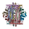
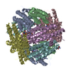
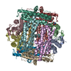
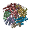
 Assembly
 Mass: 18.015 Da / Num. of mol.: 68 / Source method: isolated from a natural source / Formula: H2O
 Sample preparation
 / Beamline: X10SA / Wavelength: 0.97
  Processing