 Movie
Movie Controller

[English] 日本語
 Yorodumi
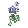
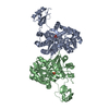
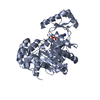
- PDB-2w6m: Crystal structure of Biotin carboxylase from E. coli in complex w... -
+ Open data

- Basic information
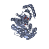
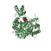
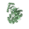
| Entry | Database: PDB / ID: 2w6m | ||||||
|---|---|---|---|---|---|---|---|
| Title | Crystal structure of Biotin carboxylase from E. coli in complex with amino-oxazole fragment series | ||||||
 Components Components | BIOTIN CARBOXYLASE | ||||||
 Keywords Keywords | LIGASE / ATP-BINDING / FATTY ACID BIOSYNTHESIS / NUCLEOTIDE-BINDING / LIPID SYNTHESIS / ATP-GRASP DOMAIN / FRAGMENT SCREENING | ||||||
| Function / homology |  Function and homology information Function and homology informationbiotin carboxylase / malonyl-CoA biosynthetic process / acetyl-CoA carboxylase complex / biotin carboxylase activity / negative regulation of fatty acid biosynthetic process / acetyl-CoA carboxylase activity / fatty acid biosynthetic process / protein homodimerization activity / ATP binding / metal ion binding ...biotin carboxylase / malonyl-CoA biosynthetic process / acetyl-CoA carboxylase complex / biotin carboxylase activity / negative regulation of fatty acid biosynthetic process / acetyl-CoA carboxylase activity / fatty acid biosynthetic process / protein homodimerization activity / ATP binding / metal ion binding / cytoplasm / cytosol Similarity search - Function | ||||||
| Biological species |  | ||||||
| Method |  X-RAY DIFFRACTION / SYNCHROTRON / MOLECULAR REPLACEMENT / Resolution: 2 Å X-RAY DIFFRACTION / SYNCHROTRON / MOLECULAR REPLACEMENT / Resolution: 2 Å | ||||||
 Authors Authors | Mochalkin, I. / Miller, J.R. | ||||||
 Citation Citation | Journal: Acs Chem.Biol. / Year: 2009 Title: Discovery of Antibacterial Biotin Carboxylase Inhibitors by Virtual Screening and Fragment-Based Approaches. Authors: Mochalkin, I. / Miller, J.R. / Narasimhan, L.S. / Thanabal, V. / Erdman, P. / Cox, P. / Prasad, J.V. / Lightle, S. / Huband, M. / Stover, K. | ||||||
| History |
|
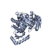
- Structure visualization
| Structure viewer | Molecule: MolmilJmol/JSmol |
|---|
- Downloads & links
-Download
| PDBx/mmCIF format | 2w6m.cif.gz | 197 KB | Display | PDBx/mmCIF format |
|---|---|---|---|---|
| PDB format | pdb2w6m.ent.gz | 156.8 KB | Display | PDB format |
| PDBx/mmJSON format | 2w6m.json.gz | Tree view | PDBx/mmJSON format | |
| Others |  Other downloads Other downloads |
-Validation report
| Arichive directory | https://data.pdbj.org/pub/pdb/validation_reports/w6/2w6mftp://data.pdbj.org/pub/pdb/validation_reports/w6/2w6m | HTTPS FTP |
|---|
-Related structure data
| Related structure data | 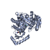 2w6nC 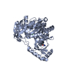 2w6oC  2w6pC 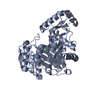 2w6qC 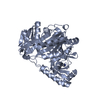 2w6zC  2w70C 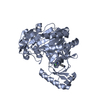 2w71C  2j9gS  2w7c S: Starting model for refinement C: citing same article ( |
|---|---|
| Similar structure data |


-Links
 PDBj
PDBj


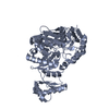
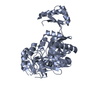
- Assembly
| Deposited unit | 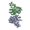
| ||||||||
|---|---|---|---|---|---|---|---|---|---|
| 1 | 
| ||||||||
| 2 | 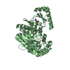
| ||||||||
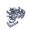
| Unit cell |
|
-Components
| #1: Protein | Mass: 49386.656 Da / Num. of mol.: 2 Source method: isolated from a genetically manipulated source Source: (gene. exp.) References: UniProt: P24182, biotin carboxylase, acetyl-CoA carboxylase #2: Chemical |   Mass: 267.079 Da / Num. of mol.: 2 / Source method: obtained synthetically / Formula: C10H7BrN2O2 Mass: 267.079 Da / Num. of mol.: 2 / Source method: obtained synthetically / Formula: C10H7BrN2O2#3: Chemical |   Mass: 35.453 Da / Num. of mol.: 2 / Source method: obtained synthetically / Formula: Cl Mass: 35.453 Da / Num. of mol.: 2 / Source method: obtained synthetically / Formula: Cl#4: Water | ChemComp-HOH / |  Mass: 18.015 Da / Num. of mol.: 810 / Source method: isolated from a natural source / Formula: H2O Mass: 18.015 Da / Num. of mol.: 810 / Source method: isolated from a natural source / Formula: H2ONonpolymer details | (2-AMINO-OXAZOL-5-YL)-(3-BROMO-PHENYL)-METHANONE (OA1): INITIAL FRAGMENT SCREENING HIT | |
|---|
-Experimental details
-Experiment
| Experiment | Method: X-RAY DIFFRACTION / Number of used crystals: 1 |
|---|
- Sample preparation
| Crystal | Density Matthews: 2.52 Å3/Da / Density % sol: 50.89 % / Description: NONE |
|---|
-Data collection
| Diffraction | Mean temperature: 100 K |
|---|---|
| Diffraction source | Source: SYNCHROTRON / Site: APS  / Beamline: 17-ID / Wavelength: 1 / Beamline: 17-ID / Wavelength: 1 |
| Detector | Type: ADSC CCD / Detector: CCD |
| Radiation | Protocol: SINGLE WAVELENGTH / Monochromatic (M) / Laue (L): M / Scattering type: x-ray |
| Radiation wavelength | Wavelength: 1 Å / Relative weight: 1 |
| Reflection | Resolution: 2→20 Å / Num. obs: 74778 / % possible obs: 99.9 % / Observed criterion σ(I): 0 / Redundancy: 6.82 % / Rmerge(I) obs: 0.08 / Net I/σ(I): 25.23 |
| Reflection shell | Resolution: 2→2.07 Å / Redundancy: 6.7 % / Rmerge(I) obs: 0.5 / Mean I/σ(I) obs: 4.47 / % possible all: 99.7 |
- Processing
| Software |
| ||||||||||||||||||||||||||||||||||||||||||||||||||||||||||||||||||||||||||||||||||||||||||||||||||||||||||||||||||||||||||||||||||||||||||||||||||||||||||||||||||||||||||||||||||||||
|---|---|---|---|---|---|---|---|---|---|---|---|---|---|---|---|---|---|---|---|---|---|---|---|---|---|---|---|---|---|---|---|---|---|---|---|---|---|---|---|---|---|---|---|---|---|---|---|---|---|---|---|---|---|---|---|---|---|---|---|---|---|---|---|---|---|---|---|---|---|---|---|---|---|---|---|---|---|---|---|---|---|---|---|---|---|---|---|---|---|---|---|---|---|---|---|---|---|---|---|---|---|---|---|---|---|---|---|---|---|---|---|---|---|---|---|---|---|---|---|---|---|---|---|---|---|---|---|---|---|---|---|---|---|---|---|---|---|---|---|---|---|---|---|---|---|---|---|---|---|---|---|---|---|---|---|---|---|---|---|---|---|---|---|---|---|---|---|---|---|---|---|---|---|---|---|---|---|---|---|---|---|---|---|
| Refinement | Method to determine structure: MOLECULAR REPLACEMENT Starting model: PDB ENTRY 2J9G Resolution: 2→80.06 Å / Cor.coef. Fo:Fc: 0.948 / Cor.coef. Fo:Fc free: 0.934 / SU B: 5.831 / SU ML: 0.089 / Cross valid method: THROUGHOUT / ESU R: 0.164 / ESU R Free: 0.14 / Stereochemistry target values: MAXIMUM LIKELIHOOD / Details: HYDROGENS HAVE BEEN ADDED IN THE RIDING POSITIONS.
| ||||||||||||||||||||||||||||||||||||||||||||||||||||||||||||||||||||||||||||||||||||||||||||||||||||||||||||||||||||||||||||||||||||||||||||||||||||||||||||||||||||||||||||||||||||||
| Solvent computation | Ion probe radii: 0.8 Å / Shrinkage radii: 0.8 Å / VDW probe radii: 1.4 Å / Solvent model: MASK | ||||||||||||||||||||||||||||||||||||||||||||||||||||||||||||||||||||||||||||||||||||||||||||||||||||||||||||||||||||||||||||||||||||||||||||||||||||||||||||||||||||||||||||||||||||||
| Displacement parameters | Biso mean: 24.87 Å2
| ||||||||||||||||||||||||||||||||||||||||||||||||||||||||||||||||||||||||||||||||||||||||||||||||||||||||||||||||||||||||||||||||||||||||||||||||||||||||||||||||||||||||||||||||||||||
| Refinement step | Cycle: LAST / Resolution: 2→80.06 Å
| ||||||||||||||||||||||||||||||||||||||||||||||||||||||||||||||||||||||||||||||||||||||||||||||||||||||||||||||||||||||||||||||||||||||||||||||||||||||||||||||||||||||||||||||||||||||
| Refine LS restraints |
|