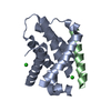
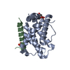
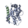
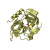
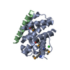
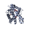
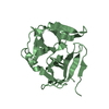
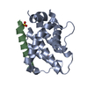
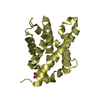
Activation, myristolyation of BID and translocation to mitochondria / positive regulation of autophagy in response to ER overload / Activation, translocation and oligomerization of BAX / Activation and oligomerization of BAK protein / Activation of BAD and translocation to mitochondria / BH3-only proteins associate with and inactivate anti-apoptotic BCL-2 members / mitochondrial outer membrane permeabilization / BH domain binding / positive regulation of fibroblast apoptotic process / B cell apoptotic process ...Activation, myristolyation of BID and translocation to mitochondria / positive regulation of autophagy in response to ER overload / Activation, translocation and oligomerization of BAX / Activation and oligomerization of BAK protein / Activation of BAD and translocation to mitochondria / BH3-only proteins associate with and inactivate anti-apoptotic BCL-2 members / mitochondrial outer membrane permeabilization / BH domain binding / positive regulation of fibroblast apoptotic process / B cell apoptotic process / negative regulation of intrinsic apoptotic signaling pathway in response to DNA damage / protein targeting to mitochondrion / regulation of epithelial cell proliferation / establishment of protein localization to membrane / negative regulation of B cell apoptotic process / positive regulation of extrinsic apoptotic signaling pathway / positive regulation of mitochondrial membrane potential / channel activity / regulation of mitochondrial membrane permeability involved in apoptotic process / mitochondrial fusion / mitochondrial ATP synthesis coupled electron transport / regulation of T cell proliferation / hepatocyte apoptotic process / apoptotic mitochondrial changes / regulation of G1/S transition of mitotic cell cycle / positive regulation of release of cytochrome c from mitochondria / B cell homeostasis / supramolecular fiber organization / positive regulation of intrinsic apoptotic signaling pathway / signal transduction in response to DNA damage / positive regulation of apoptotic signaling pathway / extrinsic apoptotic signaling pathway / extrinsic apoptotic signaling pathway in absence of ligand / release of cytochrome c from mitochondria / positive regulation of protein-containing complex assembly / mitochondrial membrane / intrinsic apoptotic signaling pathway in response to DNA damage / protein-containing complex assembly / regulation of apoptotic process / mitochondrial outer membrane / positive regulation of apoptotic process / protein heterodimerization activity / ubiquitin protein ligase binding / protein-containing complex binding / negative regulation of apoptotic process / protein homodimerization activity / mitochondrion / membrane / cytosol / cytoplasm Similarity search - Function
BH3-interacting domain death agonist / Bcl-2-related protein A1 / BH3 interacting domain (BID) / Blc2-like / Apoptosis Regulator Bcl-x / Apoptosis regulator, Bcl-2, BH3 motif, conserved site / Apoptosis regulator, Bcl-2 family BH3 motif signature. / Apoptosis regulator, Bcl-2, BH1 motif, conserved site / Apoptosis regulator, Bcl-2 family BH1 motif signature. / Apoptosis regulator, Bcl-2, BH2 motif, conserved site ...BH3-interacting domain death agonist / Bcl-2-related protein A1 / BH3 interacting domain (BID) / Blc2-like / Apoptosis Regulator Bcl-x / Apoptosis regulator, Bcl-2, BH3 motif, conserved site / Apoptosis regulator, Bcl-2 family BH3 motif signature. / Apoptosis regulator, Bcl-2, BH1 motif, conserved site / Apoptosis regulator, Bcl-2 family BH1 motif signature. / Apoptosis regulator, Bcl-2, BH2 motif, conserved site / Apoptosis regulator, Bcl-2 family BH2 motif signature. / BCL (B-Cell lymphoma); contains BH1, BH2 regions / Bcl-2 family / Bcl-2, Bcl-2 homology region 1-3 / Bcl2-like / Apoptosis regulator proteins, Bcl-2 family / BCL2-like apoptosis inhibitors family profile. / Bcl-2-like superfamily / Orthogonal Bundle / Mainly Alpha Similarity search - Domain/homology
 Movie
Movie Controller


 Open data
 Basic information
 Components
 Keywords
 APOPTOSIS / PROTEIN-PROTEIN COMPLEX / BH3 /
  Function and homology information
 Authors
 Citation
 Structure visualization
 Downloads & links
 Other downloads


 PDBj
PDBj
 Assembly


 Mass: 35.453 Da / Num. of mol.: 1 / Source method: obtained synthetically / Formula: Cl
 Mass: 18.015 Da / Num. of mol.: 25 / Source method: isolated from a natural source / Formula: H2O
 Sample preparation
 Processing