hydrolase activity, acting on glycosyl bonds / hydrolase activity, hydrolyzing O-glycosyl compounds / carbohydrate metabolic process / metal ion binding Similarity search - Function
2-ACETAMIDO-1,2-DIDEOXYNOJIRMYCIN / Alpha-N-acetylglucosaminidase family protein / Alpha-N-acetylglucosaminidase family protein Similarity search - Component
SHEET DETERMINATION METHOD: DSSP THE SHEETS PRESENTED AS "AD" IN EACH CHAIN ON SHEET RECORDS BELOW ... SHEET DETERMINATION METHOD: DSSP THE SHEETS PRESENTED AS "AD" IN EACH CHAIN ON SHEET RECORDS BELOW IS ACTUALLY AN 8-STRANDED BARREL THIS IS REPRESENTED BY A 9-STRANDED SHEET IN WHICH THE FIRST AND LAST STRANDS ARE IDENTICAL. THE SHEET STRUCTURE OF THIS MOLECULE IS BIFURCATED. IN ORDER TO REPRESENT THIS FEATURE IN THE SHEET RECORDS BELOW, TWO SHEETS ARE DEFINED.
Method to determine structure: OTHER Starting model: NONE Resolution: 2.36→19.7 Å / Cor.coef. Fo:Fc: 0.946 / Cor.coef. Fo:Fc free: 0.921 / SU B: 7.672 / SU ML: 0.183 / Cross valid method: THROUGHOUT / ESU R: 0.407 / ESU R Free: 0.259 / Stereochemistry target values: MAXIMUM LIKELIHOOD / Details: HYDROGENS HAVE BEEN ADDED IN THE RIDING POSITIONS.
Rfactor
Num. reflection
% reflection
Selection details
Rfree
0.246
2198
5 %
RANDOM
Rwork
0.203
-
-
-
obs
0.205
41573
90.7 %
-
Solvent computation
Ion probe radii: 0.8 Å / Shrinkage radii: 0.8 Å / VDW probe radii: 1.4 Å / Solvent model: MASK
 Movie
Movie Controller

 Yorodumi
 Open data
 Basic information
 Components
 Keywords
 Function and homology information
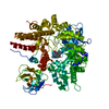
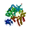
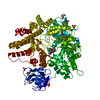
 CLOSTRIDIUM PERFRINGENS (bacteria)
 X-RAY DIFFRACTION / OTHER / Resolution: 2.36 Å
  Authors
 Citation
 Structure visualization
 Downloads & links
 Other downloads


 PDBj
PDBj


 Assembly
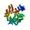
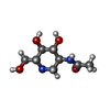
 Mass: 204.224 Da / Num. of mol.: 1 / Source method: obtained synthetically / Formula: C8H16N2O4
 Mass: 92.094 Da / Num. of mol.: 2 / Source method: obtained synthetically / Formula: C3H8O3
 Mass: 40.078 Da / Num. of mol.: 1 / Source method: obtained synthetically / Formula: Ca
 Mass: 18.015 Da / Num. of mol.: 381 / Source method: isolated from a natural source / Formula: H2O
 Sample preparation
 Processing