 Movie
Movie Controller

[English] 日本語
 Yorodumi
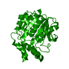
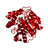
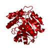
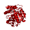
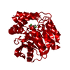
- PDB-2pu7: Crystal Structure of S112A/H265A double mutant of a C-C hydrolase... -
+ Open data

- Basic information
| Entry | Database: PDB / ID: 2pu7 | ||||||
|---|---|---|---|---|---|---|---|
| Title | Crystal Structure of S112A/H265A double mutant of a C-C hydrolase, BphD, from Burkholderia xenovorans LB400 | ||||||
 Components Components | 2-hydroxy-6-oxo-6-phenylhexa-2,4-dienoate hydrolase | ||||||
 Keywords Keywords | HYDROLASE / C-C bond hydrolase / Structural Genomics | ||||||
| Function / homology |  Function and homology information Function and homology information2-hydroxy-6-oxonona-2,4-dienedioate hydrolase activity / 2,6-dioxo-6-phenylhexa-3-enoate hydrolase / 2,6-dioxo-6-phenylhexa-3-enoate hydrolase activity / : Similarity search - Function | ||||||
| Biological species |  Burkholderia xenovorans (bacteria) Burkholderia xenovorans (bacteria) | ||||||
| Method |  X-RAY DIFFRACTION / SYNCHROTRON / MOLECULAR REPLACEMENT / Resolution: 2.07 Å X-RAY DIFFRACTION / SYNCHROTRON / MOLECULAR REPLACEMENT / Resolution: 2.07 Å | ||||||
 Authors Authors | Bhowmik, S. / Bolin, J.T. | ||||||
 Citation Citation | Journal: J.Biol.Chem. / Year: 2007 Title: The Tautomeric Half-reaction of BphD, a C-C Bond Hydrolase: KINETIC AND STRUCTURAL EVIDENCE SUPPORTING A KEY ROLE FOR HISTIDINE 265 OF THE CATALYTIC TRIAD. Authors: Horsman, G.P. / Bhowmik, S. / Seah, S.Y. / Kumar, P. / Bolin, J.T. / Eltis, L.D. | ||||||
| History |
|
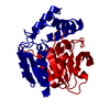
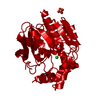
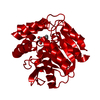
- Structure visualization
| Structure viewer | Molecule: MolmilJmol/JSmol |
|---|
- Downloads & links
-Download
| PDBx/mmCIF format | 2pu7.cif.gz | 73.3 KB | Display | PDBx/mmCIF format |
|---|---|---|---|---|
| PDB format | pdb2pu7.ent.gz | 53.3 KB | Display | PDB format |
| PDBx/mmJSON format | 2pu7.json.gz | Tree view | PDBx/mmJSON format | |
| Others |  Other downloads Other downloads |
-Validation report
| Summary document | 2pu7_validation.pdf.gz | 439.5 KB | Display | wwPDB validaton report |
|---|---|---|---|---|
| Full document | 2pu7_full_validation.pdf.gz | 440.9 KB | Display | |
| Data in XML | 2pu7_validation.xml.gz | 12.9 KB | Display | |
| Data in CIF | 2pu7_validation.cif.gz | 17.4 KB | Display | |
| Arichive directory | https://data.pdbj.org/pub/pdb/validation_reports/pu/2pu7ftp://data.pdbj.org/pub/pdb/validation_reports/pu/2pu7 | HTTPS FTP |
-Related structure data
| Related structure data | 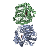 2pu5C 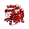 2puhC  2pujC 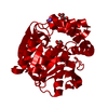 2ri6C 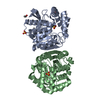 2og1S S: Starting model for refinement C: citing same article ( |
|---|---|
| Similar structure data |


-Links
 PDBj
PDBj


- Assembly
| Deposited unit | 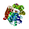
| ||||||||
|---|---|---|---|---|---|---|---|---|---|
| 1 | 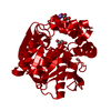
| ||||||||
| Unit cell |
| ||||||||
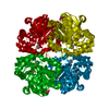
| Details | The biological assembly is a tetramer generated from the monomer in the asymmetric unit by the operations: x, y, z; -x+1/2, -y+1/2, z+1/2; -y, x+1/2, z+1/4; y+1/2, -x, z+3/4; |
-Components
| #1: Protein | Mass: 31985.582 Da / Num. of mol.: 1 / Mutation: S112A, H265A Source method: isolated from a genetically manipulated source Source: (gene. exp.) Burkholderia xenovorans (bacteria) / Strain: LB400 / Gene: bphD / Production host: | ||
|---|---|---|---|
| #2: Chemical | ChemComp-NA /   Mass: 22.990 Da / Num. of mol.: 1 / Source method: obtained synthetically / Formula: Na Mass: 22.990 Da / Num. of mol.: 1 / Source method: obtained synthetically / Formula: Na | ||
| #3: Chemical |   Mass: 102.046 Da / Num. of mol.: 2 / Source method: obtained synthetically / Formula: C3H2O4 Mass: 102.046 Da / Num. of mol.: 2 / Source method: obtained synthetically / Formula: C3H2O4#4: Water | ChemComp-HOH / |  Mass: 18.015 Da / Num. of mol.: 73 / Source method: isolated from a natural source / Formula: H2O Mass: 18.015 Da / Num. of mol.: 73 / Source method: isolated from a natural source / Formula: H2O |
-Experimental details
-Experiment
| Experiment | Method: X-RAY DIFFRACTION / Number of used crystals: 1 |
|---|
- Sample preparation
| Crystal | Density Matthews: 2.33 Å3/Da / Density % sol: 47.11 % |
|---|---|
| Crystal grow | Temperature: 293 K / Method: vapor diffusion, sitting drop / pH: 6.5 Details: 1.9 M sodium malonate, pH 6.5, VAPOR DIFFUSION, SITTING DROP, temperature 100K, temperature 293K |
-Data collection
| Diffraction | Mean temperature: 100 K |
|---|---|
| Diffraction source | Source: SYNCHROTRON / Site: APS  / Beamline: 22-ID / Wavelength: 1 Å / Beamline: 22-ID / Wavelength: 1 Å |
| Detector | Type: MARMOSAIC 300 mm CCD / Detector: CCD / Date: Jul 5, 2006 |
| Radiation | Protocol: SINGLE WAVELENGTH / Monochromatic (M) / Laue (L): M / Scattering type: x-ray |
| Radiation wavelength | Wavelength: 1 Å / Relative weight: 1 |
| Reflection | Resolution: 2.07→82.2 Å / Num. obs: 18453 / % possible obs: 98.8 % / Observed criterion σ(F): 2 / Observed criterion σ(I): 2 / Redundancy: 9.3 % / Rsym value: 8.4 / Net I/σ(I): 23.9 |
| Reflection shell | Resolution: 2.07→2.125 Å / Redundancy: 6.7 % / Mean I/σ(I) obs: 3 / Num. unique all: 1697 / Rsym value: 8.4 / % possible all: 92.4 |
- Processing
| Software |
| ||||||||||||||||||||||||||||||||||||||||||||||||||||||||||||||||||||||||||||||||||||||||||||||||||||
|---|---|---|---|---|---|---|---|---|---|---|---|---|---|---|---|---|---|---|---|---|---|---|---|---|---|---|---|---|---|---|---|---|---|---|---|---|---|---|---|---|---|---|---|---|---|---|---|---|---|---|---|---|---|---|---|---|---|---|---|---|---|---|---|---|---|---|---|---|---|---|---|---|---|---|---|---|---|---|---|---|---|---|---|---|---|---|---|---|---|---|---|---|---|---|---|---|---|---|---|---|---|
| Refinement | Method to determine structure: MOLECULAR REPLACEMENT Starting model: PDB ENTRY 2OG1 Resolution: 2.07→82.2 Å / Cor.coef. Fo:Fc: 0.952 / Cor.coef. Fo:Fc free: 0.915 / SU B: 2.714 / SU ML: 0.077 / Cross valid method: THROUGHOUT / σ(F): 0 / ESU R: 0.203 / ESU R Free: 0.2 / Stereochemistry target values: MAXIMUM LIKELIHOOD / Details: HYDROGENS HAVE BEEN ADDED IN THE RIDING POSITIONS
| ||||||||||||||||||||||||||||||||||||||||||||||||||||||||||||||||||||||||||||||||||||||||||||||||||||
| Solvent computation | Ion probe radii: 0.8 Å / Shrinkage radii: 0.8 Å / VDW probe radii: 1.4 Å / Solvent model: BABINET MODEL WITH MASK | ||||||||||||||||||||||||||||||||||||||||||||||||||||||||||||||||||||||||||||||||||||||||||||||||||||
| Displacement parameters | Biso mean: 28.257 Å2
| ||||||||||||||||||||||||||||||||||||||||||||||||||||||||||||||||||||||||||||||||||||||||||||||||||||
| Refine analyze | Luzzati coordinate error obs: 0.231 Å | ||||||||||||||||||||||||||||||||||||||||||||||||||||||||||||||||||||||||||||||||||||||||||||||||||||
| Refinement step | Cycle: LAST / Resolution: 2.07→82.2 Å
| ||||||||||||||||||||||||||||||||||||||||||||||||||||||||||||||||||||||||||||||||||||||||||||||||||||
| Refine LS restraints |
| ||||||||||||||||||||||||||||||||||||||||||||||||||||||||||||||||||||||||||||||||||||||||||||||||||||
| LS refinement shell | Resolution: 2.07→2.125 Å / Total num. of bins used: 20
|
