 Movie
Movie Controller

[English] 日本語
 Yorodumi
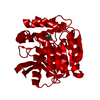
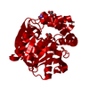
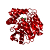
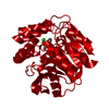
- PDB-3v1m: Crystal Structure of the S112A/H265Q mutant of a C-C hydrolase, B... -
+ Open data

- Basic information
| Entry | Database: PDB / ID: 3v1m | ||||||
|---|---|---|---|---|---|---|---|
| Title | Crystal Structure of the S112A/H265Q mutant of a C-C hydrolase, BphD from Burkholderia xenovorans LB400, after exposure to its substrate HOPDA | ||||||
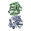
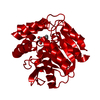
 Components Components | 2-hydroxy-6-oxo-6-phenylhexa-2,4-dienoate hydrolase | ||||||
 Keywords Keywords | HYDROLASE / C-C bond hydrolase / Alpha/beta hydrolase fold / BPHD / ALPHA/BETA HYDROLASE / PCB DEGRADATION / META CLEAVAGE PRODUCT HYDROLASE / MCP HYDROLASE / 2-HYDROXY-6-OXO-6-PHENYL-HEXA-2 / 4-DIENOATE HYDROLASE | ||||||
| Function / homology |  Function and homology information Function and homology informationbiphenyl catabolic process / 2-hydroxy-6-oxonona-2,4-dienedioate hydrolase activity / 2,6-dioxo-6-phenylhexa-3-enoate hydrolase activity / 2,6-dioxo-6-phenylhexa-3-enoate hydrolase / acylglycerol catabolic process / monoacylglycerol lipase activity / membrane Similarity search - Function | ||||||
| Biological species |  Burkholderia xenovorans (bacteria) Burkholderia xenovorans (bacteria) | ||||||
| Method |  X-RAY DIFFRACTION / SYNCHROTRON / MOLECULAR REPLACEMENT / molecular replacement / Resolution: 1.92 Å X-RAY DIFFRACTION / SYNCHROTRON / MOLECULAR REPLACEMENT / molecular replacement / Resolution: 1.92 Å | ||||||
 Authors Authors | Ghosh, S. / Bolin, J.T. | ||||||
 Citation Citation | Journal: J.Am.Chem.Soc. / Year: 2012 Title: Identification of an Acyl-Enzyme Intermediate in a meta-Cleavage Product Hydrolase Reveals the Versatility of the Catalytic Triad. Authors: Ruzzini, A.C. / Ghosh, S. / Horsman, G.P. / Foster, L.J. / Bolin, J.T. / Eltis, L.D. | ||||||
| History |
|
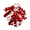
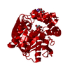
- Structure visualization
| Structure viewer | Molecule: MolmilJmol/JSmol |
|---|
- Downloads & links
-Download
| PDBx/mmCIF format | 3v1m.cif.gz | 74 KB | Display | PDBx/mmCIF format |
|---|---|---|---|---|
| PDB format | pdb3v1m.ent.gz | 54.9 KB | Display | PDB format |
| PDBx/mmJSON format | 3v1m.json.gz | Tree view | PDBx/mmJSON format | |
| Others |  Other downloads Other downloads |
-Validation report
| Arichive directory | https://data.pdbj.org/pub/pdb/validation_reports/v1/3v1mftp://data.pdbj.org/pub/pdb/validation_reports/v1/3v1m | HTTPS FTP |
|---|
-Related structure data


-Links
 PDBj
PDBj


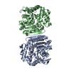
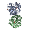
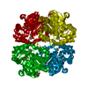
- Assembly
| Deposited unit | 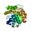
| ||||||||
|---|---|---|---|---|---|---|---|---|---|
| 1 | 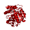
| ||||||||
| Unit cell |
|
-Components
| #1: Protein | Mass: 32042.633 Da / Num. of mol.: 1 / Mutation: S112A,H265Q Source method: isolated from a genetically manipulated source Source: (gene. exp.) Burkholderia xenovorans (bacteria) / Strain: LB400 / Gene: bphD, Bxeno_C1120, Bxe_C1186 / Production host: References: UniProt: P47229, 2,6-dioxo-6-phenylhexa-3-enoate hydrolase |
|---|---|
| #2: Chemical | ChemComp-MLI /   Mass: 102.046 Da / Num. of mol.: 1 / Source method: obtained synthetically / Formula: C3H2O4 Mass: 102.046 Da / Num. of mol.: 1 / Source method: obtained synthetically / Formula: C3H2O4 |
| #3: Chemical | ChemComp-HPK / (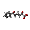  Mass: 217.197 Da / Num. of mol.: 1 / Source method: obtained synthetically / Formula: C12H9O4 Mass: 217.197 Da / Num. of mol.: 1 / Source method: obtained synthetically / Formula: C12H9O4 |
| #4: Water | ChemComp-HOH /  Mass: 18.015 Da / Num. of mol.: 121 / Source method: isolated from a natural source / Formula: H2O Mass: 18.015 Da / Num. of mol.: 121 / Source method: isolated from a natural source / Formula: H2O |
| Nonpolymer details | WATER 379 MAY BE A CATION SUCH AS NA+ BUT THIS WAS NOT TESTED |
-Experimental details
-Experiment
| Experiment | Method: X-RAY DIFFRACTION / Number of used crystals: 1 |
|---|
- Sample preparation
| Crystal | Density Matthews: 2.33 Å3/Da / Density % sol: 47.24 % |
|---|---|
| Crystal grow | Temperature: 298 K / Method: vapor diffusing, sitting drop, microseeding / pH: 7 Details: 2.4 M Sodium malonate, pH 7.0, VAPOR DIFFUSING, SITTING DROP, MICROSEEDING, temperature 298K |
-Data collection
| Diffraction | Mean temperature: 100 K | |||||||||||||||||||||||||||||||||||||||||||||||||||||||||||||||||||||||||||||
|---|---|---|---|---|---|---|---|---|---|---|---|---|---|---|---|---|---|---|---|---|---|---|---|---|---|---|---|---|---|---|---|---|---|---|---|---|---|---|---|---|---|---|---|---|---|---|---|---|---|---|---|---|---|---|---|---|---|---|---|---|---|---|---|---|---|---|---|---|---|---|---|---|---|---|---|---|---|---|
| Diffraction source | Source: SYNCHROTRON / Site: APS  / Beamline: 21-ID-F / Wavelength: 0.97872 Å / Beamline: 21-ID-F / Wavelength: 0.97872 Å | |||||||||||||||||||||||||||||||||||||||||||||||||||||||||||||||||||||||||||||
| Detector | Type: MARMOSAIC 225 mm CCD / Detector: CCD / Date: Dec 6, 2009 | |||||||||||||||||||||||||||||||||||||||||||||||||||||||||||||||||||||||||||||
| Radiation | Monochromator: C(111) / Protocol: SINGLE WAVELENGTH / Monochromatic (M) / Laue (L): M / Scattering type: x-ray | |||||||||||||||||||||||||||||||||||||||||||||||||||||||||||||||||||||||||||||
| Radiation wavelength | Wavelength: 0.97872 Å / Relative weight: 1 | |||||||||||||||||||||||||||||||||||||||||||||||||||||||||||||||||||||||||||||
| Reflection | Resolution: 1.92→82.79 Å / Num. obs: 23211 / % possible obs: 99.4 % / Redundancy: 8.1 % / Rmerge(I) obs: 0.075 / Χ2: 1.004 / Net I/σ(I): 10.7 | |||||||||||||||||||||||||||||||||||||||||||||||||||||||||||||||||||||||||||||
| Reflection shell |
|
-Phasing
| Phasing | Method: molecular replacement | |||||||||
|---|---|---|---|---|---|---|---|---|---|---|
| Phasing MR | Rfactor: 32.11 / Model details: Phaser MODE: MR_AUTO
|
- Processing
| Software |
| ||||||||||||||||||||||||||||||||||||||||||||||||||||||||||||
|---|---|---|---|---|---|---|---|---|---|---|---|---|---|---|---|---|---|---|---|---|---|---|---|---|---|---|---|---|---|---|---|---|---|---|---|---|---|---|---|---|---|---|---|---|---|---|---|---|---|---|---|---|---|---|---|---|---|---|---|---|---|
| Refinement | Method to determine structure: MOLECULAR REPLACEMENT / Resolution: 1.92→82.79 Å / Cor.coef. Fo:Fc: 0.961 / Cor.coef. Fo:Fc free: 0.937 / WRfactor Rfree: 0.227 / WRfactor Rwork: 0.1803 / Occupancy max: 1 / Occupancy min: 0.4 / FOM work R set: 0.8357 / SU B: 3.516 / SU ML: 0.103 / SU R Cruickshank DPI: 0.1593 / SU Rfree: 0.1475 / Cross valid method: THROUGHOUT / σ(F): 0 / ESU R: 0.159 / ESU R Free: 0.148 / Stereochemistry target values: MAXIMUM LIKELIHOOD Details: HYDROGENS HAVE BEEN ADDED IN THE RIDING POSITIONS U VALUES : REFINED INDIVIDUALLY
| ||||||||||||||||||||||||||||||||||||||||||||||||||||||||||||
| Solvent computation | Ion probe radii: 0.8 Å / Shrinkage radii: 0.8 Å / VDW probe radii: 1.2 Å / Solvent model: MASK | ||||||||||||||||||||||||||||||||||||||||||||||||||||||||||||
| Displacement parameters | Biso max: 57.01 Å2 / Biso mean: 24.7694 Å2 / Biso min: 14.03 Å2
| ||||||||||||||||||||||||||||||||||||||||||||||||||||||||||||
| Refinement step | Cycle: LAST / Resolution: 1.92→82.79 Å
| ||||||||||||||||||||||||||||||||||||||||||||||||||||||||||||
| Refine LS restraints |
| ||||||||||||||||||||||||||||||||||||||||||||||||||||||||||||
| LS refinement shell | Resolution: 1.922→1.972 Å / Total num. of bins used: 20
|
