 Movie
Movie Controller

[English] 日本語
 Yorodumi
- PDB-2pea: NMR Based Structure of the Closed Conformation of LYS48-Linked Di... -
+ Open data

- Basic information
| Entry | Database: PDB / ID: 2pea | ||||||
|---|---|---|---|---|---|---|---|
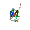
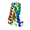
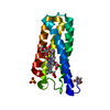
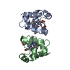
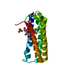
| Title | NMR Based Structure of the Closed Conformation of LYS48-Linked Di-Ubiquitin Using Experimental Global Rotational Diffusion Tensor from NMR Relaxation Measurements | ||||||
 Components Components | Ubiquitin | ||||||
 Keywords Keywords | SIGNALING PROTEIN / UBIQUITIN / LYS48-LINKED DI-UBIQUITIN / POLYUBIQUITIN | ||||||
| Function / homology |  Function and homology information Function and homology information: / : / protein modification process => GO:0036211 / Peptide chain elongation / Selenocysteine synthesis / Formation of a pool of free 40S subunits / Eukaryotic Translation Termination / SRP-dependent cotranslational protein targeting to membrane / Response of EIF2AK4 (GCN2) to amino acid deficiency / Viral mRNA Translation ...: / : / protein modification process => GO:0036211 / Peptide chain elongation / Selenocysteine synthesis / Formation of a pool of free 40S subunits / Eukaryotic Translation Termination / SRP-dependent cotranslational protein targeting to membrane / Response of EIF2AK4 (GCN2) to amino acid deficiency / Viral mRNA Translation / Nonsense Mediated Decay (NMD) independent of the Exon Junction Complex (EJC) / GTP hydrolysis and joining of the 60S ribosomal subunit / L13a-mediated translational silencing of Ceruloplasmin expression / Major pathway of rRNA processing in the nucleolus and cytosol / Nonsense Mediated Decay (NMD) enhanced by the Exon Junction Complex (EJC) / : / Maturation of protein E / Maturation of protein E / ER Quality Control Compartment (ERQC) / Myoclonic epilepsy of Lafora / FLT3 signaling by CBL mutants / Constitutive Signaling by NOTCH1 HD Domain Mutants / IRAK2 mediated activation of TAK1 complex / Prevention of phagosomal-lysosomal fusion / Alpha-protein kinase 1 signaling pathway / Glycogen synthesis / IRAK1 recruits IKK complex / IRAK1 recruits IKK complex upon TLR7/8 or 9 stimulation / Endosomal Sorting Complex Required For Transport (ESCRT) / Membrane binding and targetting of GAG proteins / Negative regulation of FLT3 / Regulation of TBK1, IKKε (IKBKE)-mediated activation of IRF3, IRF7 / PTK6 Regulates RTKs and Their Effectors AKT1 and DOK1 / Regulation of TBK1, IKKε-mediated activation of IRF3, IRF7 upon TLR3 ligation / IRAK2 mediated activation of TAK1 complex upon TLR7/8 or 9 stimulation / NOTCH2 Activation and Transmission of Signal to the Nucleus / TICAM1,TRAF6-dependent induction of TAK1 complex / TICAM1-dependent activation of IRF3/IRF7 / APC/C:Cdc20 mediated degradation of Cyclin B / Regulation of FZD by ubiquitination / Downregulation of ERBB4 signaling / cytosolic ribosome / APC-Cdc20 mediated degradation of Nek2A / p75NTR recruits signalling complexes / InlA-mediated entry of Listeria monocytogenes into host cells / TRAF6 mediated IRF7 activation in TLR7/8 or 9 signaling / Regulation of pyruvate metabolism / TRAF6-mediated induction of TAK1 complex within TLR4 complex / NF-kB is activated and signals survival / Regulation of innate immune responses to cytosolic DNA / Pexophagy / Downregulation of ERBB2:ERBB3 signaling / NRIF signals cell death from the nucleus / Activated NOTCH1 Transmits Signal to the Nucleus / Regulation of PTEN localization / VLDLR internalisation and degradation / Synthesis of active ubiquitin: roles of E1 and E2 enzymes / Translesion synthesis by REV1 / Regulation of BACH1 activity / TICAM1, RIP1-mediated IKK complex recruitment / MAP3K8 (TPL2)-dependent MAPK1/3 activation / Translesion synthesis by POLK / InlB-mediated entry of Listeria monocytogenes into host cell / Degradation of CDH1 / JNK (c-Jun kinases) phosphorylation and activation mediated by activated human TAK1 / Activation of IRF3, IRF7 mediated by TBK1, IKKε (IKBKE) / Josephin domain DUBs / Downregulation of TGF-beta receptor signaling / Translesion synthesis by POLI / Gap-filling DNA repair synthesis and ligation in GG-NER / Degradation of CRY and PER proteins / IKK complex recruitment mediated by RIP1 / Regulation of activated PAK-2p34 by proteasome mediated degradation / PINK1-PRKN Mediated Mitophagy / TGF-beta receptor signaling in EMT (epithelial to mesenchymal transition) / TNFR1-induced NF-kappa-B signaling pathway / Autodegradation of Cdh1 by Cdh1:APC/C / TCF dependent signaling in response to WNT / APC/C:Cdc20 mediated degradation of Securin / Regulation of NF-kappa B signaling / N-glycan trimming in the ER and Calnexin/Calreticulin cycle / activated TAK1 mediates p38 MAPK activation / Asymmetric localization of PCP proteins / Ubiquitin-dependent degradation of Cyclin D / SCF-beta-TrCP mediated degradation of Emi1 / NIK-->noncanonical NF-kB signaling / TNFR2 non-canonical NF-kB pathway / Regulation of signaling by CBL / AUF1 (hnRNP D0) binds and destabilizes mRNA / NOTCH3 Activation and Transmission of Signal to the Nucleus / Negative regulators of DDX58/IFIH1 signaling / Assembly of the pre-replicative complex / Vpu mediated degradation of CD4 / Negative regulation of FGFR3 signaling / Fanconi Anemia Pathway / Deactivation of the beta-catenin transactivating complex / Peroxisomal protein import / Degradation of DVL / Stabilization of p53 / Cdc20:Phospho-APC/C mediated degradation of Cyclin A Similarity search - Function | ||||||
| Biological species |  Homo sapiens (human) Homo sapiens (human) | ||||||
| Method | SOLUTION NMR | ||||||
 Authors Authors | Ryabov, Y. / Fushman, D. | ||||||
 Citation Citation | Journal: J.Am.Chem.Soc. / Year: 2007 Title: Structural assembly of multidomain proteins and protein complexes guided by the overall rotational diffusion tensor. Authors: Ryabov, Y. / Fushman, D. #1: Journal: J.Am.Chem.Soc. / Year: 2006 Title: An efficient computational method for predicting rotational diffusion tensors of globular proteins using an ellipsoid representation. Authors: Ryabov, Y. / Geraghty, C. / Varshney, A. / Fushman, D. #2: Journal: J.Am.Chem.Soc. / Year: 2007 Title: A Model of Interdomain Mobility in a Multidomain Protein Authors: Ryabov, Y. / Fushman, D. #3: Journal: J.Mol.Biol. / Year: 2002 Title: Structural Properties of Polyubiquitin Chains in Solution Authors: Varadan, R. / Walker, O. / Pickart, C. / Fushman, D. | ||||||
| History |
|
- Structure visualization
| Structure viewer | Molecule: MolmilJmol/JSmol |
|---|
- Downloads & links
-Download
| PDBx/mmCIF format | 2pea.cif.gz | 61.2 KB | Display | PDBx/mmCIF format |
|---|---|---|---|---|
| PDB format | pdb2pea.ent.gz | 45.8 KB | Display | PDB format |
| PDBx/mmJSON format | 2pea.json.gz | Tree view | PDBx/mmJSON format | |
| Others |  Other downloads Other downloads |
-Validation report
| Arichive directory | https://data.pdbj.org/pub/pdb/validation_reports/pe/2peaftp://data.pdbj.org/pub/pdb/validation_reports/pe/2pea | HTTPS FTP |
|---|
-Related structure data


-Links
 PDBj
PDBj


- Assembly
| Deposited unit | 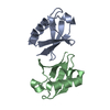
| |||||||||
|---|---|---|---|---|---|---|---|---|---|---|
| 1 |
| |||||||||
| NMR ensembles |
|
-Components
| #1: Protein | Mass: 8576.831 Da / Num. of mol.: 2 Source method: isolated from a genetically manipulated source Source: (gene. exp.) Homo sapiens (human) / Production host:  |
|---|
-Experimental details
-Experiment
| Experiment | Method: SOLUTION NMR | ||||||||||||||||||||||||||||||||
|---|---|---|---|---|---|---|---|---|---|---|---|---|---|---|---|---|---|---|---|---|---|---|---|---|---|---|---|---|---|---|---|---|---|
| NMR experiment |
|
 HSQC
HSQC- Sample preparation
| Details | Contents: DI-UBIQUITIN, 90% WATER/10% D20 |
|---|---|
| Sample conditions | Ionic strength: 20mM / pH: 6.8 / Pressure: AMBIENT / Temperature: 298.0 K |
-NMR measurement
| Radiation | Protocol: SINGLE WAVELENGTH / Monochromatic (M) / Laue (L): M |
|---|---|
| Radiation wavelength | Relative weight: 1 |
| NMR spectrometer | Type: Bruker AVANCE / Manufacturer: Bruker / Model: AVANCE / Field strength: 600 MHz |
- Processing
| NMR software |
| |||||||||
|---|---|---|---|---|---|---|---|---|---|---|
| Refinement | Software ordinal: 1 Details: THE STRUCTURE WAS DETERMINED WITH PROGRAM ELM USING COMPLETE ROTATIONAL DIFFUSION TENSOR OF DI-UBIQUITIN AS EXPERIMENTAL RESTRAINT FOR BOTH ORIENTATION AND POSITIONING OF THE INDIVIDUAL ...Details: THE STRUCTURE WAS DETERMINED WITH PROGRAM ELM USING COMPLETE ROTATIONAL DIFFUSION TENSOR OF DI-UBIQUITIN AS EXPERIMENTAL RESTRAINT FOR BOTH ORIENTATION AND POSITIONING OF THE INDIVIDUAL UBIQUITIN DOMAINS WITHIN THE MOLECULE. UBIQUITIN DOMAINS WHERE ORIENTED BY A RIGID BODY ROTATION USING EXPERIMENTALLY DERIVED PRINCIPAL AXES FRAME OF THE DIFFUSION TENSOR (SEE REFERENCE 2). THE RELATIVE DOMAIN POSITIONS IN DI-UBIQUITIN WERE DETERMINED BY A RIGID BODY TRANSLATION USING ALL COMPONENTS OF THE EXPERIMENTALLY DERIVED ROTATIONAL DIFFUSION TENSOR AS RESTRAINTS. FOR EACH UBIQUITIN DOMAIN THE STRUCTURE OF THE FIRST CONFORMER OF PDB ENTRY 1D3Z WAS ASSUMED. THE DEPOSITED CONFORMATION REPRESENTS ONE OF THE TWO EXPERIMENTALLY DETECTABLE CONFORMATIONS OF DI-UBIQUITIN AT PH 6.8. THE OCCUPATION PROBABILITY OF THIS CONFORMATION IS APPROXIMATELY 90%. CHAIN A CORRESPONDS TO UBIQUITIN DOMAIN THAT HAS A FREE C-TERMINUS IN DI-UBIQUITIN. CHAIN B CORRESPONDS TO UBIQUITIN DOMAIN THAT IN DI-UBIQUITIN IS LINKED VIA AN ISOPEPTIDE BOND BETWEEN ITS C- TERMINAL GLY76 AND LYS48 OF CHAIN A. THE ISOPEPTIDE BOND WAS PRESENT IN THE DI-UBIQUITIN MOLECULE IN THIS STUDY. HOWEVER, BECAUSE THIS STRUCTURE WAS OBTAINED BY A RIGID BODY ROTATION AND TRANSLATION AND NO CONSTRAINTS REPRESENTING THE INTERDOMAIN LINKAGE WERE INCLUDED, THE ISOPEPTIDE LINKAGE IS NOT PRESENT IN THE STRUCTURE. FLEXIBLE C-TERMINI OF BOTH UBIQUTIN DOMAINS (RESIDUES 73-76)WERE EXCLUDED FROM THE NMR RATA ANALYSIS AND THEREFORE ARE NOT PRESENT IN THIS STRUCTURE. | |||||||||
| NMR representative | Selection criteria: lowest energy | |||||||||
| NMR ensemble | Conformer selection criteria: structures with the lowest energy Conformers submitted total number: 1 |
