electron transport chain / electron transfer activity / periplasmic space / iron ion binding / heme binding Similarity search - Function
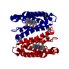
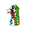
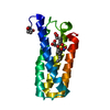
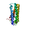
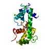
Cytochrome c prime, subgroup / Cytochrome c prime / Cytochrome c class II profile. / Cytochrome c, class II / Cytochrome C' / Cytochrome c/b562 / Cytochrome c/b562 / Four Helix Bundle (Hemerythrin (Met), subunit A) / Up-down Bundle / Mainly Alpha Similarity search - Domain/homology
Resolution: 1.06→45.24 Å / Cor.coef. Fo:Fc: 0.972 / Cor.coef. Fo:Fc free: 0.961 / SU B: 1.121 / SU ML: 0.024 / Cross valid method: THROUGHOUT / ESU R: 0.03 / ESU R Free: 0.031 / Details: HYDROGENS HAVE BEEN ADDED IN THE RIDING POSITIONS
Rfactor
Num. reflection
% reflection
Selection details
Rfree
0.20005
3506
5 %
RANDOM
Rwork
0.17758
-
-
-
obs
0.1787
66609
96.7 %
-
Solvent computation
Ion probe radii: 0.8 Å / Shrinkage radii: 0.8 Å / VDW probe radii: 1.2 Å
 Movie
Movie Controller

 Yorodumi
 Open data
 Basic information
 Components
 Keywords
 Function and homology information
 Alcaligenes xylosoxydans xylosoxydans (bacteria)
 X-RAY DIFFRACTION /
  Authors
 Citation
 Structure visualization
 Downloads & links
 Other downloads
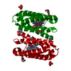


 PDBj
PDBj


 Assembly
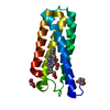

 Mass: 618.503 Da / Num. of mol.: 1 / Source method: obtained synthetically / Formula: C34H34FeN4O4
 Mass: 46.005 Da / Num. of mol.: 1 / Source method: obtained synthetically / Formula: NO2
 Mass: 92.094 Da / Num. of mol.: 1 / Source method: obtained synthetically / Formula: C3H8O3
 Mass: 18.015 Da / Num. of mol.: 214 / Source method: isolated from a natural source / Formula: H2O
 Sample preparation
 / Beamline: I04-1 / Wavelength: 0.9282 Å
 Processing