 Movie
Movie Controller

[English] 日本語
 Yorodumi
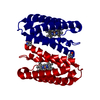
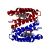
- PDB-5nc0: The 0.91 A resolution structure of the L16G mutant of cytochrome ... -
+ Open data

- Basic information
| Entry | Database: PDB / ID: 5nc0 | |||||||||
|---|---|---|---|---|---|---|---|---|---|---|
| Title | The 0.91 A resolution structure of the L16G mutant of cytochrome c prime from Alcaligenes xylosoxidans, complexed with nitric oxide | |||||||||
 Components Components | Cytochrome c' | |||||||||
 Keywords Keywords | OXIDOREDUCTASE / nitric oxide / cytochrome / homodimer | |||||||||
| Function / homology |  Function and homology information Function and homology informationelectron transport chain / electron transfer activity / periplasmic space / iron ion binding / heme binding Similarity search - Function | |||||||||
| Biological species |  Alcaligenes xylosoxydans xylosoxydans (bacteria) Alcaligenes xylosoxydans xylosoxydans (bacteria) | |||||||||
| Method |  X-RAY DIFFRACTION / SYNCHROTRON / MOLECULAR REPLACEMENT / Resolution: 0.91 Å X-RAY DIFFRACTION / SYNCHROTRON / MOLECULAR REPLACEMENT / Resolution: 0.91 Å | |||||||||
 Authors Authors | Strange, R. / Hough, M. / Antonyuk, S. / Rustage, N. | |||||||||
 Citation Citation | Journal: Inorg Chem / Year: 2017 Title: Distinguishing Nitro vs Nitrito Coordination in Cytochrome c' Using Vibrational Spectroscopy and Density Functional Theory. Authors: Nilsson, Z.N. / Mandella, B.L. / Sen, K. / Kekilli, D. / Hough, M.A. / Moenne-Loccoz, P. / Strange, R.W. / Andrew, C.R. | |||||||||
| History |
|
- Structure visualization
| Structure viewer | Molecule: MolmilJmol/JSmol |
|---|
- Downloads & links
-Download
| PDBx/mmCIF format | 5nc0.cif.gz | 80 KB | Display | PDBx/mmCIF format |
|---|---|---|---|---|
| PDB format | pdb5nc0.ent.gz | 58.3 KB | Display | PDB format |
| PDBx/mmJSON format | 5nc0.json.gz | Tree view | PDBx/mmJSON format | |
| Others |  Other downloads Other downloads |
-Validation report
| Arichive directory | https://data.pdbj.org/pub/pdb/validation_reports/nc/5nc0ftp://data.pdbj.org/pub/pdb/validation_reports/nc/5nc0 | HTTPS FTP |
|---|
-Related structure data
| Related structure data | 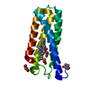 5ngxC 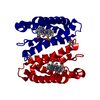 2yl7S S: Starting model for refinement C: citing same article ( |
|---|---|
| Similar structure data |


-Links
 PDBj
PDBj


- Assembly
| Deposited unit | 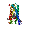
| ||||||||
|---|---|---|---|---|---|---|---|---|---|
| 1 | 
| ||||||||
| Unit cell |
|
-Components
-Protein , 1 types, 1 molecules A
| #1: Protein | Mass: 13575.336 Da / Num. of mol.: 1 / Mutation: L16G Source method: isolated from a genetically manipulated source Details: residue PCA is pyroglutamic acid - 2-pyrrolidone-5-carboxylic acid Source: (gene. exp.) Alcaligenes xylosoxydans xylosoxydans (bacteria)Production host: |
|---|
-Non-polymers , 7 types, 242 molecules 


| #2: Chemical | ChemComp-HEC /  Mass: 618.503 Da / Num. of mol.: 1 / Source method: obtained synthetically / Formula: C34H34FeN4O4 Mass: 618.503 Da / Num. of mol.: 1 / Source method: obtained synthetically / Formula: C34H34FeN4O4 | ||||||
|---|---|---|---|---|---|---|---|
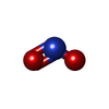
| #3: Chemical | ChemComp-NO /  Mass: 30.006 Da / Num. of mol.: 1 / Source method: obtained synthetically / Formula: NO Mass: 30.006 Da / Num. of mol.: 1 / Source method: obtained synthetically / Formula: NO | ||||||
| #4: Chemical | ChemComp-NO3 /  Mass: 62.005 Da / Num. of mol.: 1 / Source method: obtained synthetically / Formula: NO3 Mass: 62.005 Da / Num. of mol.: 1 / Source method: obtained synthetically / Formula: NO3 | ||||||
| #5: Chemical |  Mass: 46.005 Da / Num. of mol.: 2 / Source method: obtained synthetically / Formula: NO2 Mass: 46.005 Da / Num. of mol.: 2 / Source method: obtained synthetically / Formula: NO2#6: Chemical | ChemComp-SO4 /  Mass: 96.063 Da / Num. of mol.: 4 / Source method: obtained synthetically / Formula: SO4 Mass: 96.063 Da / Num. of mol.: 4 / Source method: obtained synthetically / Formula: SO4#7: Chemical |  Mass: 62.068 Da / Num. of mol.: 3 / Source method: obtained synthetically / Formula: C2H6O2 Mass: 62.068 Da / Num. of mol.: 3 / Source method: obtained synthetically / Formula: C2H6O2#8: Water | ChemComp-HOH / | Mass: 18.015 Da / Num. of mol.: 230 / Source method: isolated from a natural source / Formula: H2O |
-Details
| Has protein modification | Y |
|---|
-Experimental details
-Experiment
| Experiment | Method: X-RAY DIFFRACTION / Number of used crystals: 1 |
|---|
- Sample preparation
| Crystal | Density Matthews: 2.67 Å3/Da / Density % sol: 53.88 % |
|---|---|
| Crystal grow | Temperature: 298 K / Method: vapor diffusion, hanging drop / pH: 7.5 / Details: AMMONIUM SULPHATE, TRIS PH 7.5 |
-Data collection
| Diffraction | Mean temperature: 100 K |
|---|---|
| Diffraction source | Source: SYNCHROTRON / Site: SOLEIL  / Beamline: PROXIMA 1 / Wavelength: 0.7872 Å / Beamline: PROXIMA 1 / Wavelength: 0.7872 Å |
| Detector | Type: ADSC QUANTUM 315r / Detector: CCD / Date: Sep 5, 2010 |
| Radiation | Protocol: SINGLE WAVELENGTH / Monochromatic (M) / Laue (L): M / Scattering type: x-ray |
| Radiation wavelength | Wavelength: 0.7872 Å / Relative weight: 1 |
| Reflection | Resolution: 0.91→44.07 Å / Num. obs: 106845 / % possible obs: 99.5 % / Redundancy: 8 % / CC1/2: 0.999 / Rpim(I) all: 0.023 / Net I/σ(I): 16.1 |
| Reflection shell | Resolution: 0.91→0.93 Å / Redundancy: 4.4 % / Mean I/σ(I) obs: 1.3 / Num. unique obs: 4889 / CC1/2: 0.547 / Rpim(I) all: 0.564 / % possible all: 93.9 |
- Processing
| Software |
| ||||||||||||||||||||||||||||||||||||||||||||||||||||||||||||||||||||||||||||||||||||||||||||||||||||||||||||||||||||||||||||||||||||||||||||||||||||||||||||||||||||||||||||||||||||||
|---|---|---|---|---|---|---|---|---|---|---|---|---|---|---|---|---|---|---|---|---|---|---|---|---|---|---|---|---|---|---|---|---|---|---|---|---|---|---|---|---|---|---|---|---|---|---|---|---|---|---|---|---|---|---|---|---|---|---|---|---|---|---|---|---|---|---|---|---|---|---|---|---|---|---|---|---|---|---|---|---|---|---|---|---|---|---|---|---|---|---|---|---|---|---|---|---|---|---|---|---|---|---|---|---|---|---|---|---|---|---|---|---|---|---|---|---|---|---|---|---|---|---|---|---|---|---|---|---|---|---|---|---|---|---|---|---|---|---|---|---|---|---|---|---|---|---|---|---|---|---|---|---|---|---|---|---|---|---|---|---|---|---|---|---|---|---|---|---|---|---|---|---|---|---|---|---|---|---|---|---|---|---|---|
| Refinement | Method to determine structure: MOLECULAR REPLACEMENT Starting model: 2YL7 Resolution: 0.91→44.07 Å / Cor.coef. Fo:Fc: 0.979 / Cor.coef. Fo:Fc free: 0.978 / SU B: 0.378 / SU ML: 0.01 / Cross valid method: THROUGHOUT / ESU R: 0.014 / ESU R Free: 0.014 / Details: HYDROGENS HAVE BEEN ADDED IN THE RIDING POSITIONS
| ||||||||||||||||||||||||||||||||||||||||||||||||||||||||||||||||||||||||||||||||||||||||||||||||||||||||||||||||||||||||||||||||||||||||||||||||||||||||||||||||||||||||||||||||||||||
| Solvent computation | Ion probe radii: 0.8 Å / Shrinkage radii: 0.8 Å / VDW probe radii: 1.2 Å | ||||||||||||||||||||||||||||||||||||||||||||||||||||||||||||||||||||||||||||||||||||||||||||||||||||||||||||||||||||||||||||||||||||||||||||||||||||||||||||||||||||||||||||||||||||||
| Displacement parameters | Biso mean: 8.977 Å2
| ||||||||||||||||||||||||||||||||||||||||||||||||||||||||||||||||||||||||||||||||||||||||||||||||||||||||||||||||||||||||||||||||||||||||||||||||||||||||||||||||||||||||||||||||||||||
| Refinement step | Cycle: 1 / Resolution: 0.91→44.07 Å
| ||||||||||||||||||||||||||||||||||||||||||||||||||||||||||||||||||||||||||||||||||||||||||||||||||||||||||||||||||||||||||||||||||||||||||||||||||||||||||||||||||||||||||||||||||||||
| Refine LS restraints |
|