 Movie
Movie Controller

[English] 日本語
 Yorodumi
- PDB-2mro: Structure of the complex of ubiquitin and the UBA domain from DNA... -
+ Open data

- Basic information
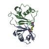
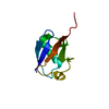
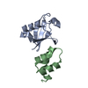
| Entry | Database: PDB / ID: 2mro | ||||||
|---|---|---|---|---|---|---|---|
| Title | Structure of the complex of ubiquitin and the UBA domain from DNA-damage-inducible 1 protein (Ddi1) | ||||||
 Components Components |
| ||||||
 Keywords Keywords | TRANSPORT PROTEIN/SIGNALING PROTEIN / DNA-damage-inducible 1 protein / Ubiquitin associated domain / Ddi1 / UBA / HYDROLASE-SIGNALING PROTEIN complex / TRANSPORT PROTEIN-SIGNALING PROTEIN complex | ||||||
| Function / homology |  Function and homology information Function and homology informationproteasome regulatory particle binding / protein transport to vacuole involved in ubiquitin-dependent protein catabolic process via the multivesicular body sorting pathway / hypothalamus gonadotrophin-releasing hormone neuron development / female meiosis I / positive regulation of protein monoubiquitination / fat pad development / mitochondrion transport along microtubule / Hydrolases; Acting on peptide bonds (peptidases); Aspartic endopeptidases / female gonad development / seminiferous tubule development ...proteasome regulatory particle binding / protein transport to vacuole involved in ubiquitin-dependent protein catabolic process via the multivesicular body sorting pathway / hypothalamus gonadotrophin-releasing hormone neuron development / female meiosis I / positive regulation of protein monoubiquitination / fat pad development / mitochondrion transport along microtubule / Hydrolases; Acting on peptide bonds (peptidases); Aspartic endopeptidases / female gonad development / seminiferous tubule development / male meiosis I / protein secretion / polyubiquitin modification-dependent protein binding / positive regulation of intrinsic apoptotic signaling pathway by p53 class mediator / energy homeostasis / neuron projection morphogenesis / vesicle-mediated transport / regulation of proteasomal protein catabolic process / Maturation of protein E / Maturation of protein E / ER Quality Control Compartment (ERQC) / Myoclonic epilepsy of Lafora / FLT3 signaling by CBL mutants / Constitutive Signaling by NOTCH1 HD Domain Mutants / IRAK2 mediated activation of TAK1 complex / Prevention of phagosomal-lysosomal fusion / Alpha-protein kinase 1 signaling pathway / Glycogen synthesis / IRAK1 recruits IKK complex / IRAK1 recruits IKK complex upon TLR7/8 or 9 stimulation / Endosomal Sorting Complex Required For Transport (ESCRT) / Membrane binding and targetting of GAG proteins / Negative regulation of FLT3 / Regulation of TBK1, IKKε (IKBKE)-mediated activation of IRF3, IRF7 / PTK6 Regulates RTKs and Their Effectors AKT1 and DOK1 / Regulation of TBK1, IKKε-mediated activation of IRF3, IRF7 upon TLR3 ligation / IRAK2 mediated activation of TAK1 complex upon TLR7/8 or 9 stimulation / NOTCH2 Activation and Transmission of Signal to the Nucleus / TICAM1,TRAF6-dependent induction of TAK1 complex / TICAM1-dependent activation of IRF3/IRF7 / APC/C:Cdc20 mediated degradation of Cyclin B / Regulation of FZD by ubiquitination / Downregulation of ERBB4 signaling / APC-Cdc20 mediated degradation of Nek2A / p75NTR recruits signalling complexes / InlA-mediated entry of Listeria monocytogenes into host cells / TRAF6 mediated IRF7 activation in TLR7/8 or 9 signaling / Regulation of pyruvate metabolism / TRAF6-mediated induction of TAK1 complex within TLR4 complex / regulation of neuron apoptotic process / NF-kB is activated and signals survival / Regulation of innate immune responses to cytosolic DNA / Pexophagy / Downregulation of ERBB2:ERBB3 signaling / NRIF signals cell death from the nucleus / Activated NOTCH1 Transmits Signal to the Nucleus / Regulation of PTEN localization / VLDLR internalisation and degradation / positive regulation of DNA replication / Synthesis of active ubiquitin: roles of E1 and E2 enzymes / ubiquitin binding / Translesion synthesis by REV1 / Regulation of BACH1 activity / TICAM1, RIP1-mediated IKK complex recruitment / positive regulation of protein ubiquitination / MAP3K8 (TPL2)-dependent MAPK1/3 activation / Translesion synthesis by POLK / InlB-mediated entry of Listeria monocytogenes into host cell / Degradation of CDH1 / JNK (c-Jun kinases) phosphorylation and activation mediated by activated human TAK1 / Activation of IRF3, IRF7 mediated by TBK1, IKKε (IKBKE) / Josephin domain DUBs / Downregulation of TGF-beta receptor signaling / Translesion synthesis by POLI / Gap-filling DNA repair synthesis and ligation in GG-NER / Degradation of CRY and PER proteins / IKK complex recruitment mediated by RIP1 / SNARE binding / Regulation of activated PAK-2p34 by proteasome mediated degradation / PINK1-PRKN Mediated Mitophagy / TGF-beta receptor signaling in EMT (epithelial to mesenchymal transition) / regulation of mitochondrial membrane potential / TNFR1-induced NF-kappa-B signaling pathway / Autodegradation of Cdh1 by Cdh1:APC/C / TCF dependent signaling in response to WNT / APC/C:Cdc20 mediated degradation of Securin / Regulation of NF-kappa B signaling / N-glycan trimming in the ER and Calnexin/Calreticulin cycle / activated TAK1 mediates p38 MAPK activation / Asymmetric localization of PCP proteins / Ubiquitin-dependent degradation of Cyclin D / SCF-beta-TrCP mediated degradation of Emi1 / NIK-->noncanonical NF-kB signaling / TNFR2 non-canonical NF-kB pathway / Regulation of signaling by CBL / AUF1 (hnRNP D0) binds and destabilizes mRNA / NOTCH3 Activation and Transmission of Signal to the Nucleus / Negative regulators of DDX58/IFIH1 signaling / Assembly of the pre-replicative complex / Vpu mediated degradation of CD4 Similarity search - Function | ||||||
| Biological species |  Homo sapiens (human) Homo sapiens (human) | ||||||
| Method | SOLUTION NMR / simulated annealing | ||||||
| Model details | lowest energy, model1 | ||||||
 Authors Authors | Zhang, D. / Fushman, D. | ||||||
 Citation Citation | Journal: Structure / Year: 2015 Title: DNA-Damage-Inducible 1 Protein (Ddi1) Contains an Uncharacteristic Ubiquitin-like Domain that Binds Ubiquitin. Authors: Nowicka, U. / Zhang, D. / Walker, O. / Krutauz, D. / Castaneda, C.A. / Chaturvedi, A. / Chen, T.Y. / Reis, N. / Glickman, M.H. / Fushman, D. | ||||||
| History |
|
- Structure visualization
| Structure viewer | Molecule: MolmilJmol/JSmol |
|---|
- Downloads & links
-Download
| PDBx/mmCIF format | 2mro.cif.gz | 258.3 KB | Display | PDBx/mmCIF format |
|---|---|---|---|---|
| PDB format | pdb2mro.ent.gz | 216.7 KB | Display | PDB format |
| PDBx/mmJSON format | 2mro.json.gz | Tree view | PDBx/mmJSON format | |
| Others |  Other downloads Other downloads |
-Validation report
| Arichive directory | https://data.pdbj.org/pub/pdb/validation_reports/mr/2mroftp://data.pdbj.org/pub/pdb/validation_reports/mr/2mro | HTTPS FTP |
|---|
-Related structure data


-Links
 PDBj
PDBj


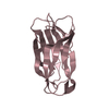
- Assembly
| Deposited unit | 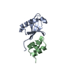
| |||||||||
|---|---|---|---|---|---|---|---|---|---|---|
| 1 |
| |||||||||
| NMR ensembles |
|
-Components
| #1: Protein | Mass: 8576.831 Da / Num. of mol.: 1 / Fragment: Human Ubiquitin Source method: isolated from a genetically manipulated source Source: (gene. exp.) Homo sapiens (human) / Gene: UBB / Production host:  |
|---|---|
| #2: Protein/peptide | Mass: 4745.367 Da / Num. of mol.: 1 / Fragment: Ubiquitin associated domain, UNP residues 389-428 Source method: isolated from a genetically manipulated source Source: (gene. exp.) Strain: ATCC 204508 / S288c / Gene: DDI1, VSM1, YER143W / Production host: |
-Experimental details
-Experiment
| Experiment | Method: SOLUTION NMR Details: Complex structure based on chemical shift perturbation data and spin-labeling data, calculated using Haddock program | ||||||||||||
|---|---|---|---|---|---|---|---|---|---|---|---|---|---|
| NMR experiment |
| ||||||||||||
| NMR details | Text: Two titration experiments were conducted. Basically unlabeled ligand protein was added stepwise to N15-Labeled protein solution. |
 HSQC
HSQC- Sample preparation
| Details |
| ||||||||||||||||||||||||||||||||||||
|---|---|---|---|---|---|---|---|---|---|---|---|---|---|---|---|---|---|---|---|---|---|---|---|---|---|---|---|---|---|---|---|---|---|---|---|---|---|
| Sample |
| ||||||||||||||||||||||||||||||||||||
| Sample conditions | Ionic strength: 20 / pH: 6.8 / Pressure: ambient / Temperature: 298.2 K |
-NMR measurement
| NMR spectrometer | Type: Bruker Avance / Manufacturer: Bruker / Model: AVANCE / Field strength: 600 MHz |
|---|
- Processing
| NMR software |
| ||||||||||||||||||||||||||||
|---|---|---|---|---|---|---|---|---|---|---|---|---|---|---|---|---|---|---|---|---|---|---|---|---|---|---|---|---|---|
| Refinement | Method: simulated annealing / Software ordinal: 1 | ||||||||||||||||||||||||||||
| NMR representative | Selection criteria: lowest energy | ||||||||||||||||||||||||||||
| NMR ensemble | Conformer selection criteria: structures with the least restraint violations Conformers calculated total number: 10 / Conformers submitted total number: 10 |
