 Movie
Movie Controller

[English] 日本語
 Yorodumi
- PDB-2hk1: Crystal structure of D-psicose 3-epimerase (DPEase) in the presen... -
+ Open data

- Basic information
| Entry | Database: PDB / ID: 2hk1 | ||||||
|---|---|---|---|---|---|---|---|
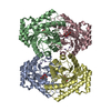
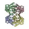
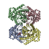
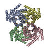
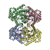
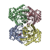
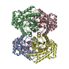
| Title | Crystal structure of D-psicose 3-epimerase (DPEase) in the presence of D-fructose | ||||||
 Components Components | D-PSICOSE 3-EPIMERASE | ||||||
 Keywords Keywords |  ISOMERASE / TIM-barrel ISOMERASE / TIM-barrel | ||||||
| Function / homology |  Function and homology information Function and homology informationD-psicose 3-epimerase / racemase and epimerase activity, acting on carbohydrates and derivatives / cobalt ion binding / manganese ion binding Similarity search - Function | ||||||
| Biological species |  Agrobacterium tumefaciens (bacteria) Agrobacterium tumefaciens (bacteria) | ||||||
| Method | X-RAY DIFFRACTION / SYNCHROTRON / MOLECULAR REPLACEMENT / Resolution: 2.3 Å | ||||||
 Authors Authors | Kim, K. / Kim, H.J. / Oh, D.K. / Cha, S.S. / Rhee, S. | ||||||
 Citation Citation | Journal: J.Mol.Biol. / Year: 2006 Title: Crystal Structure of d-Psicose 3-epimerase from Agrobacterium tumefaciens and its Complex with True Substrate d-Fructose: A Pivotal Role of Metal in Catalysis, an Active Site for the Non- ...Title: Crystal Structure of d-Psicose 3-epimerase from Agrobacterium tumefaciens and its Complex with True Substrate d-Fructose: A Pivotal Role of Metal in Catalysis, an Active Site for the Non-phosphorylated Substrate, and its Conformational Changes Authors: Kim, K. / Kim, H.J. / Oh, D.K. / Cha, S.S. / Rhee, S. | ||||||
| History |
|
- Structure visualization
| Structure viewer | Molecule: MolmilJmol/JSmol |
|---|
- Downloads & links
-Download
| PDBx/mmCIF format | 2hk1.cif.gz | 231.6 KB | Display | PDBx/mmCIF format |
|---|---|---|---|---|
| PDB format | pdb2hk1.ent.gz | 196.4 KB | Display | PDB format |
| PDBx/mmJSON format | 2hk1.json.gz | Tree view | PDBx/mmJSON format | |
| Others |  Other downloads Other downloads |
-Validation report
| Arichive directory | https://data.pdbj.org/pub/pdb/validation_reports/hk/2hk1ftp://data.pdbj.org/pub/pdb/validation_reports/hk/2hk1 | HTTPS FTP |
|---|
-Related structure data


-Links
 PDBj
PDBj
- Assembly
| Deposited unit | 
| ||||||||
|---|---|---|---|---|---|---|---|---|---|
| 1 |
| ||||||||
| Unit cell |
| ||||||||
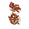
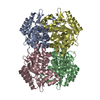
| Details | The biological assembly is a tetramer, which is generated from subunits A-D to B-C by crystallographic symmetric operation. |
-Components
| #1: Protein | Mass: 34210.301 Da / Num. of mol.: 4 Source method: isolated from a genetically manipulated source Source: (gene. exp.) Agrobacterium tumefaciens (bacteria) / Plasmid: pET15b / Production host: Escherichia coli (E. coli)References: UniProt: A9CH28, Isomerases; Intramolecular oxidoreductases; Interconverting aldoses and ketoses, and related compounds#2: Sugar | ChemComp-FUD / Fructose  Type: D-saccharide / Mass: 180.156 Da / Num. of mol.: 4 Type: D-saccharide / Mass: 180.156 Da / Num. of mol.: 4Source method: isolated from a genetically manipulated source Formula: C6H12O6 #3: Chemical | ChemComp-MN /   Mass: 54.938 Da / Num. of mol.: 4 / Source method: obtained synthetically / Formula: Mn Mass: 54.938 Da / Num. of mol.: 4 / Source method: obtained synthetically / Formula: Mn#4: Water | ChemComp-HOH / | Water Mass: 18.015 Da / Num. of mol.: 376 / Source method: isolated from a natural source / Formula: H2O Mass: 18.015 Da / Num. of mol.: 376 / Source method: isolated from a natural source / Formula: H2O |
|---|
-Experimental details
-Experiment
| Experiment | Method: X-RAY DIFFRACTION / Number of used crystals: 1 |
|---|
- Sample preparation
| Crystal | Density Matthews: 3.09 Å3/Da / Density % sol: 60.22 % |
|---|---|
| Crystal grow | Temperature: 295 K / Method: vapor diffusion, hanging drop / pH: 8 Details: 14%(w/v) PEG 1000, 0.1M imidazole (pH 8.0), 0.2M calcium acetate, VAPOR DIFFUSION, HANGING DROP, temperature 295.0K |
-Data collection
| Diffraction | Mean temperature: 297 K |
|---|---|
| Diffraction source | Source: SYNCHROTRON / Site: PAL/PLS  / Beamline: 4A / Wavelength: 0.97948 Å / Beamline: 4A / Wavelength: 0.97948 Å |
| Detector | Type: BRUKER PROTEUM 300 / Detector: CCD / Date: Jun 22, 2005 |
| Radiation | Monochromator: Si 111 CHANNEL / Protocol: SINGLE WAVELENGTH / Monochromatic (M) / Laue (L): M / Scattering type: x-ray |
| Radiation wavelength | Wavelength: 0.97948 Å / Relative weight: 1 |
| Reflection | Resolution: 2.3→50 Å / Num. obs: 69649 / % possible obs: 100 % / Observed criterion σ(F): 0 / Observed criterion σ(I): 0 / Redundancy: 6.2 % / Rmerge(I) obs: 0.069 / Χ2: 1.019 / Net I/σ(I): 10.5 |
| Reflection shell | Resolution: 2.3→2.38 Å / Redundancy: 6.2 % / Num. unique all: 6855 / Χ2: 0.869 / % possible all: 100 |
- Processing
| Software |
| ||||||||||||||||||||||||||||||||
|---|---|---|---|---|---|---|---|---|---|---|---|---|---|---|---|---|---|---|---|---|---|---|---|---|---|---|---|---|---|---|---|---|---|
| Refinement | Method to determine structure: MOLECULAR REPLACEMENT / Resolution: 2.3→33.5 Å / σ(F): 0 / Stereochemistry target values: Engh & Huber
| ||||||||||||||||||||||||||||||||
| Solvent computation | Bsol: 32.378 Å2 | ||||||||||||||||||||||||||||||||
| Displacement parameters | Biso mean: 41.938 Å2
| ||||||||||||||||||||||||||||||||
| Refinement step | Cycle: LAST / Resolution: 2.3→33.5 Å
| ||||||||||||||||||||||||||||||||
| Refine LS restraints |
| ||||||||||||||||||||||||||||||||
| Xplor file |
|
