 Movie
Movie Controller

[English] 日本語
 Yorodumi
- PDB-7bp1: Crystal structure of 2, 3-dihydroxybenzoic acid decarboxylase fro... -
+ Open data

- Basic information
| Entry | Database: PDB / ID: 7bp1 | ||||||
|---|---|---|---|---|---|---|---|
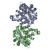
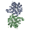
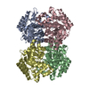
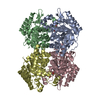
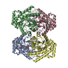
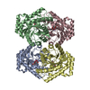
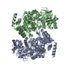
| Title | Crystal structure of 2, 3-dihydroxybenzoic acid decarboxylase from Fusarium oxysporum in complex with Catechol | ||||||
 Components Components | 2,3-dihydroxybenzoate decarboxylase | ||||||
 Keywords Keywords | BIOSYNTHETIC PROTEIN / 2 / 3-dihydroxybenzoic acid decarboxylase | ||||||
| Function / homology |  Function and homology information Function and homology informationsecondary metabolic process / carboxy-lyase activity / hydrolase activity / metal ion binding / cytosol Similarity search - Function | ||||||
| Biological species |   Fusarium oxysporum (fungus) Fusarium oxysporum (fungus) | ||||||
| Method |  X-RAY DIFFRACTION / SYNCHROTRON / MOLECULAR REPLACEMENT / Resolution: 1.97 Å X-RAY DIFFRACTION / SYNCHROTRON / MOLECULAR REPLACEMENT / Resolution: 1.97 Å | ||||||
 Authors Authors | Song, M.K. / Feng, J.H. / Liu, W.D. / Wu, Q.Q. / Zhu, D.M. | ||||||
 Citation Citation | Journal: Chembiochem / Year: 2020 Title: 2,3-Dihydroxybenzoic Acid Decarboxylase from Fusarium oxysporum: Crystal Structures and Substrate Recognition Mechanism. Authors: Song, M. / Zhang, X. / Liu, W. / Feng, J. / Cui, Y. / Yao, P. / Wang, M. / Guo, R.T. / Wu, Q. / Zhu, D. | ||||||
| History |
|
- Structure visualization
| Structure viewer | Molecule: MolmilJmol/JSmol |
|---|
- Downloads & links
-Download
| PDBx/mmCIF format | 7bp1.cif.gz | 297.4 KB | Display | PDBx/mmCIF format |
|---|---|---|---|---|
| PDB format | pdb7bp1.ent.gz | 239 KB | Display | PDB format |
| PDBx/mmJSON format | 7bp1.json.gz | Tree view | PDBx/mmJSON format | |
| Others |  Other downloads Other downloads |
-Validation report
| Arichive directory | https://data.pdbj.org/pub/pdb/validation_reports/bp/7bp1ftp://data.pdbj.org/pub/pdb/validation_reports/bp/7bp1 | HTTPS FTP |
|---|
-Related structure data
| Related structure data |  6m53C  7bpcC  2dvtS S: Starting model for refinement C: citing same article ( |
|---|---|
| Similar structure data |
-Links
 PDBj
PDBj


- Assembly
| Deposited unit | 
| |||||||||||||||||||||||||||||||||||||||||||||
|---|---|---|---|---|---|---|---|---|---|---|---|---|---|---|---|---|---|---|---|---|---|---|---|---|---|---|---|---|---|---|---|---|---|---|---|---|---|---|---|---|---|---|---|---|---|---|
| 1 |
| |||||||||||||||||||||||||||||||||||||||||||||
| Unit cell |
| |||||||||||||||||||||||||||||||||||||||||||||
| Noncrystallographic symmetry (NCS) | NCS domain:
NCS domain segments:
|
-Components
| #1: Protein | Mass: 39290.387 Da / Num. of mol.: 4 Source method: isolated from a genetically manipulated source Source: (gene. exp.) Fusarium oxysporum (fungus) / Gene: BFJ70_g2310 / Production host:  #2: Chemical |   Mass: 110.111 Da / Num. of mol.: 3 / Source method: obtained synthetically / Formula: C6H6O2 Mass: 110.111 Da / Num. of mol.: 3 / Source method: obtained synthetically / Formula: C6H6O2#3: Chemical | ChemComp-ZN / |   Mass: 65.409 Da / Num. of mol.: 1 / Source method: isolated from a natural source / Formula: Zn / Feature type: SUBJECT OF INVESTIGATION Mass: 65.409 Da / Num. of mol.: 1 / Source method: isolated from a natural source / Formula: Zn / Feature type: SUBJECT OF INVESTIGATION#4: Water | ChemComp-HOH / |  Mass: 18.015 Da / Num. of mol.: 1181 / Source method: isolated from a natural source / Formula: H2O Mass: 18.015 Da / Num. of mol.: 1181 / Source method: isolated from a natural source / Formula: H2OHas ligand of interest | Y | |
|---|
-Experimental details
-Experiment
| Experiment | Method: X-RAY DIFFRACTION / Number of used crystals: 1 |
|---|
- Sample preparation
| Crystal | Density Matthews: 2.28 Å3/Da / Density % sol: 45.96 % |
|---|---|
| Crystal grow | Temperature: 295 K / Method: vapor diffusion, sitting drop Details: 0.1 mol/L tri-potassium citrate, 20% (w/v) PEG 3350 and 10% (v/v) glycerol |
-Data collection
| Diffraction | Mean temperature: 100 K / Serial crystal experiment: N |
|---|---|
| Diffraction source | Source: SYNCHROTRON / Site: SSRF  / Beamline: BL18U1 / Wavelength: 1 Å / Beamline: BL18U1 / Wavelength: 1 Å |
| Detector | Type: DECTRIS PILATUS3 S 6M / Detector: PIXEL / Date: Oct 29, 2019 |
| Radiation | Protocol: SINGLE WAVELENGTH / Monochromatic (M) / Laue (L): M / Scattering type: x-ray |
| Radiation wavelength | Wavelength: 1 Å / Relative weight: 1 |
| Reflection | Resolution: 1.97→25 Å / Num. obs: 102008 / % possible obs: 99.7 % / Redundancy: 5.8 % / Rmerge(I) obs: 0.077 / Net I/σ(I): 11.8 |
| Reflection shell | Resolution: 1.97→2.04 Å / Redundancy: 5.2 % / Rmerge(I) obs: 0.609 / Num. unique obs: 9853 / % possible all: 98.1 |
- Processing
| Software |
| |||||||||||||||||||||||||||||||||||
|---|---|---|---|---|---|---|---|---|---|---|---|---|---|---|---|---|---|---|---|---|---|---|---|---|---|---|---|---|---|---|---|---|---|---|---|---|
| Refinement | Method to determine structure: MOLECULAR REPLACEMENT Starting model: 2DVT Resolution: 1.97→24.87 Å / SU ML: 0.2 / Cross valid method: THROUGHOUT / σ(F): 1.35 / Phase error: 20.07 / Stereochemistry target values: ML
| |||||||||||||||||||||||||||||||||||
| Solvent computation | Shrinkage radii: 0.9 Å / VDW probe radii: 1.11 Å / Solvent model: FLAT BULK SOLVENT MODEL | |||||||||||||||||||||||||||||||||||
| Displacement parameters | Biso max: 86.43 Å2 / Biso mean: 30.08 Å2 / Biso min: 15.74 Å2 | |||||||||||||||||||||||||||||||||||
| Refinement step | Cycle: final / Resolution: 1.97→24.87 Å
| |||||||||||||||||||||||||||||||||||
| Refine LS restraints NCS |
| |||||||||||||||||||||||||||||||||||
| LS refinement shell | Resolution: 1.97→2.04 Å
|
