 Movie
Movie Controller

[English] 日本語
 Yorodumi
- PDB-3vnj: Crystal structures of D-Psicose 3-epimerase with D-psicose from C... -
+ Open data

- Basic information
| Entry | Database: PDB / ID: 3vnj | ||||||
|---|---|---|---|---|---|---|---|
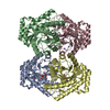
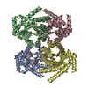
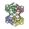
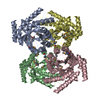
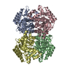
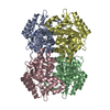
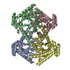
| Title | Crystal structures of D-Psicose 3-epimerase with D-psicose from Clostridium cellulolyticum H10 | ||||||
 Components Components | Xylose isomerase domain protein TIM barrel | ||||||
 Keywords Keywords | ISOMERASE / D-Psicose 3-epimerase / ketohexose | ||||||
| Function / homology |  Function and homology information Function and homology informationD-psicose 3-epimerase / racemase and epimerase activity, acting on carbohydrates and derivatives / cobalt ion binding / manganese ion binding Similarity search - Function | ||||||
| Biological species |  Clostridium cellulolyticum (bacteria) Clostridium cellulolyticum (bacteria) | ||||||
| Method |  X-RAY DIFFRACTION / SYNCHROTRON / MOLECULAR REPLACEMENT / Resolution: 2.08 Å X-RAY DIFFRACTION / SYNCHROTRON / MOLECULAR REPLACEMENT / Resolution: 2.08 Å | ||||||
 Authors Authors | Chan, H.C. / Zhu, Y. / Hu, Y. / Ko, T.P. / Huang, C.H. / Ren, F. / Chen, C.C. / Guo, R.T. / Sun, Y. | ||||||
 Citation Citation | Journal: Protein Cell / Year: 2012 Title: Crystal structures of D-psicose 3-epimerase from Clostridium cellulolyticum H10 and its complex with ketohexose sugars. Authors: Chan, H.C. / Zhu, Y. / Hu, Y. / Ko, T.P. / Huang, C.H. / Ren, F. / Chen, C.C. / Ma, Y. / Guo, R.T. / Sun, Y. | ||||||
| History |
|
- Structure visualization
| Structure viewer | Molecule: MolmilJmol/JSmol |
|---|
- Downloads & links
-Download
| PDBx/mmCIF format | 3vnj.cif.gz | 253.4 KB | Display | PDBx/mmCIF format |
|---|---|---|---|---|
| PDB format | pdb3vnj.ent.gz | 202.8 KB | Display | PDB format |
| PDBx/mmJSON format | 3vnj.json.gz | Tree view | PDBx/mmJSON format | |
| Others |  Other downloads Other downloads |
-Validation report
| Arichive directory | https://data.pdbj.org/pub/pdb/validation_reports/vn/3vnjftp://data.pdbj.org/pub/pdb/validation_reports/vn/3vnj | HTTPS FTP |
|---|
-Related structure data
| Related structure data | 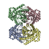 3vniSC  3vnkC 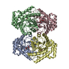 3vnlC 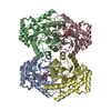 3vnmC S: Starting model for refinement C: citing same article ( |
|---|---|
| Similar structure data |


-Links
 PDBj
PDBj
- Assembly
| Deposited unit | 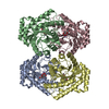
| ||||||||
|---|---|---|---|---|---|---|---|---|---|
| 1 |
| ||||||||
| Unit cell |
|
-Components
| #1: Protein | Mass: 33147.410 Da / Num. of mol.: 4 Source method: isolated from a genetically manipulated source Source: (gene. exp.) Clostridium cellulolyticum (bacteria) / Strain: H10 / Gene: Ccel_0941 / Plasmid: pET-21d / Production host: #2: Sugar | ChemComp-PSJ /   Type: D-saccharide / Mass: 180.156 Da / Num. of mol.: 4 Type: D-saccharide / Mass: 180.156 Da / Num. of mol.: 4Source method: isolated from a genetically manipulated source Formula: C6H12O6 #3: Chemical | ChemComp-MN /   Mass: 54.938 Da / Num. of mol.: 4 / Source method: obtained synthetically / Formula: Mn Mass: 54.938 Da / Num. of mol.: 4 / Source method: obtained synthetically / Formula: Mn#4: Water | ChemComp-HOH / |  Mass: 18.015 Da / Num. of mol.: 832 / Source method: isolated from a natural source / Formula: H2O Mass: 18.015 Da / Num. of mol.: 832 / Source method: isolated from a natural source / Formula: H2O |
|---|
-Experimental details
-Experiment
| Experiment | Method: X-RAY DIFFRACTION / Number of used crystals: 1 |
|---|
- Sample preparation
| Crystal | Density Matthews: 3.08 Å3/Da / Density % sol: 60 % |
|---|---|
| Crystal grow | Temperature: 298 K / Method: vapor diffusion, sitting drop / pH: 8.2 Details: 0.2M tri-sodium citrate dehydrate, 17-20% Polyethylene Glycol 3350, pH 8.2, VAPOR DIFFUSION, SITTING DROP, temperature 298K |
-Data collection
| Diffraction | Mean temperature: 100 K |
|---|---|
| Diffraction source | Source: SYNCHROTRON / Site: NSRRC  / Beamline: BL13C1 / Wavelength: 1 Å / Beamline: BL13C1 / Wavelength: 1 Å |
| Detector | Type: ADSC QUANTUM 315r / Detector: CCD / Date: Jun 26, 2011 |
| Radiation | Protocol: SINGLE WAVELENGTH / Monochromatic (M) / Laue (L): M / Scattering type: x-ray |
| Radiation wavelength | Wavelength: 1 Å / Relative weight: 1 |
| Reflection | Resolution: 2.08→25 Å / Num. obs: 93519 / % possible obs: 100 % / Observed criterion σ(I): 2 / Rsym value: 0.055 / Net I/σ(I): 25.3 |
| Reflection shell | Resolution: 2.08→2.15 Å / Redundancy: 3.6 % / Mean I/σ(I) obs: 25.26 / Num. unique all: 9224 / Rsym value: 0.48 / % possible all: 96.6 |
- Processing
| Software |
| ||||||||||||||||||
|---|---|---|---|---|---|---|---|---|---|---|---|---|---|---|---|---|---|---|---|
| Refinement | Method to determine structure: MOLECULAR REPLACEMENT Starting model: 3VNI Resolution: 2.08→25 Å / σ(F): 2
| ||||||||||||||||||
| Refinement step | Cycle: LAST / Resolution: 2.08→25 Å
| ||||||||||||||||||
| LS refinement shell | Resolution: 2.08→2.15 Å /
|
