[English] 日本語
Yorodumi
- PDB-2hk0: Crystal structure of D-psicose 3-epimerase (DPEase) in the absenc... -

+
Open data


ID or keywords:

Loading...

-
Basic information

Entry
Database: PDB / ID: 2hk0
Title
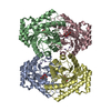
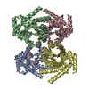
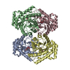
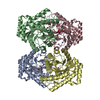
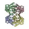
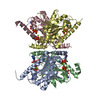
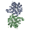
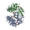
Crystal structure of D-psicose 3-epimerase (DPEase) in the absence of substrate
ComponentsD-PSICOSE 3-EPIMERASE
KeywordsISOMERASE / TIM-barrel
Function / homology
Function and homology information


D-psicose 3-epimerase / racemase and epimerase activity, acting on carbohydrates and derivatives / cobalt ion binding / manganese ion binding
Similarity search - Function
Divalent-metal-dependent TIM barrel enzymes / Xylose isomerase-like, TIM barrel domain / Xylose isomerase-like TIM barrel / Xylose isomerase-like superfamily / TIM Barrel / Alpha-Beta Barrel / Alpha Beta
Similarity search - Domain/homology
D-psicose 3-epimerase / :
Similarity search - Component
Biological speciesAgrobacterium tumefaciens (bacteria)
MethodX-RAY DIFFRACTION / SYNCHROTRON / MAD / Resolution: 2 Å
AuthorsKim, K. / Kim, H.J. / Oh, D.K. / Cha, S.S. / Rhee, S.
CitationJournal: J.Mol.Biol. / Year: 2006
Title: Crystal Structure of d-Psicose 3-epimerase from Agrobacterium tumefaciens and its Complex with True Substrate d-Fructose: A Pivotal Role of Metal in Catalysis, an Active Site for the Non- ...Title: Crystal Structure of d-Psicose 3-epimerase from Agrobacterium tumefaciens and its Complex with True Substrate d-Fructose: A Pivotal Role of Metal in Catalysis, an Active Site for the Non-phosphorylated Substrate, and its Conformational Changes
Authors: Kim, K. / Kim, H.J. / Oh, D.K. / Cha, S.S. / Rhee, S.
History
DepositionJul 3, 2006Deposition site: RCSB / Processing site: PDBJ
Revision 1.0Aug 29, 2006Provider: repository / Type: Initial release
Revision 1.1May 1, 2008Group: Version format compliance
Revision 1.2Jul 13, 2011Group: Version format compliance
Revision 1.3Oct 18, 2017Group: Refinement description / Category: software

-
Structure visualization

Structure viewerMolecule:
MolmilJmol/JSmol

Downloads & links

-
Assembly

Deposited unit
A: D-PSICOSE 3-EPIMERASE
B: D-PSICOSE 3-EPIMERASE
C: D-PSICOSE 3-EPIMERASE
D: D-PSICOSE 3-EPIMERASE


Theoretical massNumber of molelcules
Total (without water)136,8414
Polymers136,8414
Non-polymers00
Water13,673759
1


  • Idetical with deposited unit
  • defined by author&software
TypeNameSymmetry operationNumber
identity operation1_555x,y,z1
Buried area8500 Å2
ΔGint-22 kcal/mol
Surface area40680 Å2
MethodPISA
Unit cell
Length a, b, c (Å)102.400, 113.000, 131.800
Angle α, β, γ (deg.)90.000, 90.000, 90.000
Int Tables number19
Space group name H-MP212121
DetailsThe biological assembly is a tetramer, which is generated from subunits A-D to B-C by crystallographic symmetric operation.

-
Components

#1: Protein
D-PSICOSE 3-EPIMERASE / D-tagatose 3-epimerase / DTEASE / AGR_L_260p


Mass: 34210.301 Da / Num. of mol.: 4
Source method: isolated from a genetically manipulated source
Source: (gene. exp.) Agrobacterium tumefaciens (bacteria) / Production host: Escherichia coli (E. coli)
References: UniProt: Q8U6Q7, UniProt: A9CH28*PLUS, Isomerases; Intramolecular oxidoreductases; Interconverting aldoses and ketoses, and related compounds
#2: Water ChemComp-HOH / water


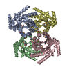
Mass: 18.015 Da / Num. of mol.: 759 / Source method: isolated from a natural source / Formula: H2O

-
Experimental details

-
Experiment

ExperimentMethod: X-RAY DIFFRACTION / Number of used crystals: 1

-
Sample preparation

CrystalDensity Matthews: 2.99 Å3/Da / Density % sol: 58.84 %
Crystal growTemperature: 295 K / Method: vapor diffusion, hanging drop / pH: 8
Details: 14%(w/v) PEG 1000, 0.1M imidazole (pH 8.0), 0.2M calcium acetate, VAPOR DIFFUSION, HANGING DROP, temperature 295.0K

-
Data collection

Diffraction
IDMean temperature (K)Crystal-ID
12971
21
31
Diffraction sourceSource: SYNCHROTRON / Site: PAL/PLS / Beamline: 6B / Wavelength: 0.97900, 0.97913, 0.97133
DetectorType: BRUKER PROTEUM 300 / Detector: CCD / Date: May 16, 2005
RadiationMonochromator: Si 111 CHANNEL / Protocol: MAD / Monochromatic (M) / Laue (L): M / Scattering type: x-ray
Radiation wavelength
IDWavelength (Å)Relative weight
10.9791
20.979131
30.971331
Reflection

D res high: 2 Å / D res low: 30 Å

IDAv σ(I) over netINumberRmerge(I) obsΧ2Num. obs% possible obs
18.214385920.1360.979993699.6
28.39692550.1191.0110257599.4
37.29679170.126110322799.4
Diffraction reflection shell
Highest resolution (Å)Lowest resolution (Å)% possible obs (%)IDRmerge(I) obsChi squared
4.313099.910.0662.059
3.424.3110010.0711.27
2.993.4299.910.0981.151
2.712.9999.710.1480.968
2.522.7199.710.2120.825
2.372.5299.510.2770.756
2.252.3799.510.360.657
2.152.2599.310.470.625
2.072.1599.210.6460.662
22.079910.8740.626
4.313010020.0561.86
3.424.3199.920.0631.261
2.993.4299.820.0921.189
2.712.9999.720.1471.009
2.522.7199.520.2170.9
2.372.5299.420.2930.824
2.252.3799.220.3970.747
2.152.2599.320.5370.713
2.072.1598.920.7410.769
22.0798.720.705
4.313010030.0481.292
3.424.3199.930.0631.133
2.993.4299.830.1041.146
2.712.9999.730.1891.032
2.522.7199.530.2990.936
2.372.5299.430.4320.907
2.252.3799.230.6140.877
2.152.2599.230.8460.852
2.072.1598.930.895
22.0797.930.858
ReflectionResolution: 2→30 Å / Num. obs: 99936 / % possible obs: 99.6 % / Observed criterion σ(F): 0 / Observed criterion σ(I): 0 / Redundancy: 14.4 % / Rmerge(I) obs: 0.098 / Χ2: 0.968
Reflection shellResolution: 2→2.07 Å / Rmerge(I) obs: 0.849 / Num. unique all: 9800 / Χ2: 0.626 / % possible all: 99

-
Phasing

PhasingMethod: MAD
Phasing set
ID
1
2
3
Phasing MADD res high: 2.2 Å / D res low: 30 Å / FOM : 0.52 / Reflection: 74053
Phasing MAD set
Clust-IDExpt-IDSet-IDWavelength (Å)F double prime refinedF prime refined
13 wavelength10.9794.6-10.21
13 wavelength20.97913.1-8.5
13 wavelength30.97132.28-4.92
Phasing MAD set site
IDAtom type symbolB isoFract xFract yFract zOccupancy
1Se28.9670.8180.5740.0740.833
2Se25.8240.790.7960.0210.969
3Se26.7050.8080.8310.1910.95
4Se27.5420.8730.7260.1870.861
5Se39.8420.9050.8330.0411.073
6Se7.6490.0430.5760.2070.665
7Se22.7170.870.7040.0450.661
8Se28.7410.7280.7450.1760.713
9Se25.8080.0030.0360.230.853
10Se25.2990.2180.2470.2190.756
11Se34.5750.0210.5140.0020.825
12Se29.2160.8070.4890.0340.868
13Se34.0490.8830.8950.2030.722
14Se26.1270.9220.7230.0110.716
15Se18.9090.970.670.1820.664
16Se9.6810.9550.5010.0630.597
17Se29.3980.6960.7570.0090.802
18Se31.6780.8870.6030.070.819
19Se30.5880.1110.6970.2240.883
20Se20.070.8270.6940.2210.613
21Se32.9140.8710.4970.1360.815
22Se37.2930.3970.7330.0590.864
23Se24.5930.0250.7120.1750.711
24Se600.0960.6330.1271.123
Phasing MAD shell
Resolution (Å)FOM Reflection
7.85-300.863848
4.98-7.850.836463
3.9-4.980.788187
3.31-3.90.79505
2.92-3.310.5710532
2.65-2.920.4211284
2.44-2.650.3211902
2.27-2.440.2112332
Phasing dmFOM : 0.71 / FOM acentric: 0.71 / FOM centric: 0.72 / Reflection: 74054 / Reflection acentric: 68588 / Reflection centric: 5466
Phasing dm shell
Resolution (Å)FOM FOM acentricFOM centricReflectionReflection acentricReflection centric
6.3-29.5620.950.960.9235392816723
3.9-6.30.930.940.871049293101182
3.1-3.90.890.90.812945119011044
2.8-3.10.770.780.711270311856847
2.4-2.80.60.60.5421601204921109
2.2-2.40.410.410.41277412213561

-
Processing

Software
NameVersionClassificationNB
DENZOdata reduction
SCALEPACKdata scaling
SOLVE2.08phasing
RESOLVE2.08phasing
CNSrefinement
PDB_EXTRACT2data extraction
HKL-2000data reduction
HKL-2000data scaling
RefinementMethod to determine structure: MAD / Resolution: 2→30 Å / σ(F): 0 / Stereochemistry target values: Engh & Huber
RfactorNum. reflection% reflectionSelection details
Rfree0.233 9704 9.4 %Random
Rwork0.197 ---
all0.197 103700 --
obs0.197 97182 93.7 %-
Solvent computationBsol: 42.556 Å2
Displacement parametersBiso mean: 26.404 Å2
Baniso -1Baniso -2Baniso -3
1--6.869 Å20 Å20 Å2
2--3.343 Å20 Å2
3---3.526 Å2
Refinement stepCycle: LAST / Resolution: 2→30 Å
ProteinNucleic acidLigandSolventTotal
Num. atoms8916 0 0 759 9675
Refine LS restraints
Refine-IDTypeDev idealDev ideal target
X-RAY DIFFRACTIONc_mcbond_it1.3731.5
X-RAY DIFFRACTIONc_scbond_it2.3922
X-RAY DIFFRACTIONc_mcangle_it1.9632
X-RAY DIFFRACTIONc_scangle_it3.3812.5
Xplor file
Refine-IDSerial noParam file
X-RAY DIFFRACTION1protein_rep.param
X-RAY DIFFRACTION2water_rep.param

+
About Yorodumi

-
News

-
Feb 9, 2022. New format data for meta-information of EMDB entries

New format data for meta-information of EMDB entries

  • Version 3 of the EMDB header file is now the official format.
  • The previous official version 1.9 will be removed from the archive.

Related info.:EMDB header

External links:wwPDB to switch to version 3 of the EMDB data model

-
Aug 12, 2020. Covid-19 info

Covid-19 info

URL: https://pdbjlvh1.pdbj.org/emnavi/covid19.php

New page: Covid-19 featured information page in EM Navigator.

Related info.:Covid-19 info / Mar 5, 2020. Novel coronavirus structure data

+
Mar 5, 2020. Novel coronavirus structure data

Novel coronavirus structure data

Related info.:Yorodumi Speices / Aug 12, 2020. Covid-19 info

External links:COVID-19 featured content - PDBj / Molecule of the Month (242):Coronavirus Proteases

+
Jan 31, 2019. EMDB accession codes are about to change! (news from PDBe EMDB page)

EMDB accession codes are about to change! (news from PDBe EMDB page)

  • The allocation of 4 digits for EMDB accession codes will soon come to an end. Whilst these codes will remain in use, new EMDB accession codes will include an additional digit and will expand incrementally as the available range of codes is exhausted. The current 4-digit format prefixed with “EMD-” (i.e. EMD-XXXX) will advance to a 5-digit format (i.e. EMD-XXXXX), and so on. It is currently estimated that the 4-digit codes will be depleted around Spring 2019, at which point the 5-digit format will come into force.
  • The EM Navigator/Yorodumi systems omit the EMD- prefix.

Related info.:Q: What is EMD? / ID/Accession-code notation in Yorodumi/EM Navigator

External links:EMDB Accession Codes are Changing Soon! / Contact to PDBj

+
Jul 12, 2017. Major update of PDB

Major update of PDB

  • wwPDB released updated PDB data conforming to the new PDBx/mmCIF dictionary.
  • This is a major update changing the version number from 4 to 5, and with Remediation, in which all the entries are updated.
  • In this update, many items about electron microscopy experimental information are reorganized (e.g. em_software).
  • Now, EM Navigator and Yorodumi are based on the updated data.

External links:wwPDB Remediation / Enriched Model Files Conforming to OneDep Data Standards Now Available in the PDB FTP Archive

-
Yorodumi

Thousand views of thousand structures

  • Yorodumi is a browser for structure data from EMDB, PDB, SASBDB, etc.
  • This page is also the successor to EM Navigator detail page, and also detail information page/front-end page for Omokage search.
  • The word "yorodu" (or yorozu) is an old Japanese word meaning "ten thousand". "mi" (miru) is to see.

Related info.:EMDB / PDB / SASBDB / Comparison of 3 databanks / Yorodumi Search / Aug 31, 2016. New EM Navigator & Yorodumi / Yorodumi Papers / Jmol/JSmol / Function and homology information / Changes in new EM Navigator and Yorodumi

Read more