 Movie
Movie Controller

[English] 日本語
 Yorodumi
- PDB-2fs6: Crystal Structure of Apo-Cellular Retinoic Acid Binding Protein T... -
+ Open data

- Basic information
| Entry | Database: PDB / ID: 2fs6 | ||||||
|---|---|---|---|---|---|---|---|
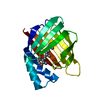
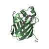
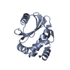
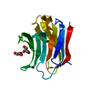
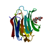
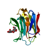
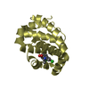
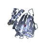
| Title | Crystal Structure of Apo-Cellular Retinoic Acid Binding Protein Type II At 1.35 Angstroms Resolution | ||||||
 Components Components | Cellular retinoic acid-binding protein 2 | ||||||
 Keywords Keywords | TRANSPORT PROTEIN / CRABPII / Retinoic Acid / Retinoids / Beta Barrel / High Resolution | ||||||
| Function / homology |  Function and homology information Function and homology informationpositive regulation of collateral sprouting / retinoid binding / retinoic acid binding / retinal binding / embryonic forelimb morphogenesis / retinoic acid metabolic process / retinol binding / Signaling by Retinoic Acid / epidermis development / fatty acid transport ...positive regulation of collateral sprouting / retinoid binding / retinoic acid binding / retinal binding / embryonic forelimb morphogenesis / retinoic acid metabolic process / retinol binding / Signaling by Retinoic Acid / epidermis development / fatty acid transport / cyclin binding / fatty acid binding / regulation of DNA-templated transcription / endoplasmic reticulum / signal transduction / extracellular exosome / nucleoplasm / nucleus / cytoplasm / cytosol Similarity search - Function | ||||||
| Biological species |  Homo sapiens (human) Homo sapiens (human) | ||||||
| Method |  X-RAY DIFFRACTION / SYNCHROTRON / Rigid body refinement / Resolution: 1.35 Å X-RAY DIFFRACTION / SYNCHROTRON / Rigid body refinement / Resolution: 1.35 Å | ||||||
 Authors Authors | Vaezeslami, S. / Geiger, J.H. | ||||||
 Citation Citation | Journal: J.Mol.Biol. / Year: 2006 Title: The structure of Apo-wild-type cellular retinoic acid binding protein II at 1.4 A and its relationship to ligand binding and nuclear translocation. Authors: Vaezeslami, S. / Mathes, E. / Vasileiou, C. / Borhan, B. / Geiger, J.H. #1: Journal: ThesisTitle: Determining crystal structures of proteins and protein complexes by X-ray crystallography: X-ray crystallographic studies of the mutants of cellular retinoic acid binding protein type II ...Title: Determining crystal structures of proteins and protein complexes by X-ray crystallography: X-ray crystallographic studies of the mutants of cellular retinoic acid binding protein type II toward designing a mimic of rhodopsin Authors: Vaezeslami, S. | ||||||
| History |
|
- Structure visualization
| Structure viewer | Molecule: MolmilJmol/JSmol |
|---|
- Downloads & links
-Download
| PDBx/mmCIF format | 2fs6.cif.gz | 139.3 KB | Display | PDBx/mmCIF format |
|---|---|---|---|---|
| PDB format | pdb2fs6.ent.gz | 109.4 KB | Display | PDB format |
| PDBx/mmJSON format | 2fs6.json.gz | Tree view | PDBx/mmJSON format | |
| Others |  Other downloads Other downloads |
-Validation report
| Arichive directory | https://data.pdbj.org/pub/pdb/validation_reports/fs/2fs6ftp://data.pdbj.org/pub/pdb/validation_reports/fs/2fs6 | HTTPS FTP |
|---|
-Related structure data
| Related structure data | 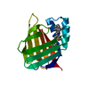 2fr3C  2frsC  2fs7C  2frr 2fru 2fs0 C: citing same article ( |
|---|---|
| Similar structure data |


-Links
 PDBj
PDBj


- Assembly
| Deposited unit | 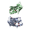
| ||||||||
|---|---|---|---|---|---|---|---|---|---|
| 1 | 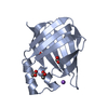
| ||||||||
| 2 | 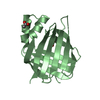
| ||||||||
| Unit cell |
|
-Components
| #1: Protein | Mass: 15581.802 Da / Num. of mol.: 2 Source method: isolated from a genetically manipulated source Source: (gene. exp.) Homo sapiens (human) / Gene: CRABP2 / Plasmid: pET17-b / Production host:  #2: Chemical | ChemComp-ACT /   Mass: 59.044 Da / Num. of mol.: 7 / Source method: obtained synthetically / Formula: C2H3O2 Mass: 59.044 Da / Num. of mol.: 7 / Source method: obtained synthetically / Formula: C2H3O2#3: Chemical | ChemComp-NA / |   Mass: 22.990 Da / Num. of mol.: 1 / Source method: obtained synthetically / Formula: Na Mass: 22.990 Da / Num. of mol.: 1 / Source method: obtained synthetically / Formula: Na#4: Chemical | ChemComp-CL / |   Mass: 35.453 Da / Num. of mol.: 1 / Source method: obtained synthetically / Formula: Cl Mass: 35.453 Da / Num. of mol.: 1 / Source method: obtained synthetically / Formula: Cl#5: Water | ChemComp-HOH / |  Mass: 18.015 Da / Num. of mol.: 391 / Source method: isolated from a natural source / Formula: H2O Mass: 18.015 Da / Num. of mol.: 391 / Source method: isolated from a natural source / Formula: H2O |
|---|
-Experimental details
-Experiment
| Experiment | Method: X-RAY DIFFRACTION / Number of used crystals: 1 |
|---|
- Sample preparation
| Crystal | Density Matthews: 2.15 Å3/Da / Density % sol: 42.71 % |
|---|---|
| Crystal grow | Temperature: 298 K / Method: vapor diffusion, hanging drop / pH: 8 Details: 30% (w/v) PEG 8000 (4000), 0.1 M TRIS-HCl pH 8.0 (8.5), and 0.2 M Sodium Acetate, vapor diffusion, hanging drop, temperature 298K |
-Data collection
| Diffraction | Mean temperature: 77 K |
|---|---|
| Diffraction source | Source: SYNCHROTRON / Site: APS  / Beamline: 14-ID-B / Wavelength: 1 Å / Beamline: 14-ID-B / Wavelength: 1 Å |
| Detector | Type: MARRESEARCH / Detector: CCD / Date: Apr 1, 2005 |
| Radiation | Protocol: SINGLE WAVELENGTH / Monochromatic (M) / Laue (L): M / Scattering type: x-ray |
| Radiation wavelength | Wavelength: 1 Å / Relative weight: 1 |
| Reflection | Resolution: 1.35→50 Å / Num. all: 56961 / Num. obs: 50380 / % possible obs: 88.4 % / Observed criterion σ(I): -3 / Redundancy: 2 % / Biso Wilson estimate: 15.4 Å2 / Rmerge(I) obs: 0.042 / Χ2: 0.591 / Net I/σ(I): 14.56 |
| Reflection shell | Resolution: 1.35→1.4 Å / % possible obs: 56.5 % / Redundancy: 1.8 % / Rmerge(I) obs: 0.261 / Mean I/σ(I) obs: 1.77 / Num. unique obs: 3219 / Χ2: 0.517 / % possible all: 56.5 |
- Processing
| Software |
| |||||||||||||||||||||||||||||||||||||||||||||||||||||||||||||||||||||||||||||||||||||||||||||||||||||||||||||||||||||||||||||||||||||||||||||||||
|---|---|---|---|---|---|---|---|---|---|---|---|---|---|---|---|---|---|---|---|---|---|---|---|---|---|---|---|---|---|---|---|---|---|---|---|---|---|---|---|---|---|---|---|---|---|---|---|---|---|---|---|---|---|---|---|---|---|---|---|---|---|---|---|---|---|---|---|---|---|---|---|---|---|---|---|---|---|---|---|---|---|---|---|---|---|---|---|---|---|---|---|---|---|---|---|---|---|---|---|---|---|---|---|---|---|---|---|---|---|---|---|---|---|---|---|---|---|---|---|---|---|---|---|---|---|---|---|---|---|---|---|---|---|---|---|---|---|---|---|---|---|---|---|---|---|---|
| Refinement | Method to determine structure: Rigid body refinement / Resolution: 1.35→17.24 Å / Cor.coef. Fo:Fc: 0.976 / Cor.coef. Fo:Fc free: 0.958 / SU B: 2.636 / SU ML: 0.047 / Cross valid method: THROUGHOUT / σ(F): 0 / ESU R: 0.076 / ESU R Free: 0.072 / Stereochemistry target values: MAXIMUM LIKELIHOOD / Details: HYDROGENS HAVE BEEN ADDED IN THE RIDING POSITIONS
| |||||||||||||||||||||||||||||||||||||||||||||||||||||||||||||||||||||||||||||||||||||||||||||||||||||||||||||||||||||||||||||||||||||||||||||||||
| Solvent computation | Ion probe radii: 0.8 Å / Shrinkage radii: 0.8 Å / VDW probe radii: 1.2 Å / Solvent model: BABINET MODEL WITH MASK | |||||||||||||||||||||||||||||||||||||||||||||||||||||||||||||||||||||||||||||||||||||||||||||||||||||||||||||||||||||||||||||||||||||||||||||||||
| Displacement parameters | Biso mean: 16.794 Å2
| |||||||||||||||||||||||||||||||||||||||||||||||||||||||||||||||||||||||||||||||||||||||||||||||||||||||||||||||||||||||||||||||||||||||||||||||||
| Refinement step | Cycle: LAST / Resolution: 1.35→17.24 Å
| |||||||||||||||||||||||||||||||||||||||||||||||||||||||||||||||||||||||||||||||||||||||||||||||||||||||||||||||||||||||||||||||||||||||||||||||||
| Refine LS restraints |
| |||||||||||||||||||||||||||||||||||||||||||||||||||||||||||||||||||||||||||||||||||||||||||||||||||||||||||||||||||||||||||||||||||||||||||||||||
| LS refinement shell | Resolution: 1.35→1.385 Å / Total num. of bins used: 20
|
