 Movie
Movie Controller

[English] 日本語
 Yorodumi
- PDB-7jwd: Cellular retinol-binding protein 2 (CRBP2) in complex with 2-lino... -
+ Open data

- Basic information
| Entry | Database: PDB / ID: 7jwd | |||||||||
|---|---|---|---|---|---|---|---|---|---|---|
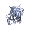
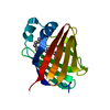
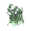
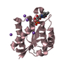
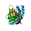
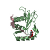
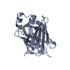
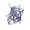
| Title | Cellular retinol-binding protein 2 (CRBP2) in complex with 2-linoleoylglycerol | |||||||||
 Components Components | Retinol-binding protein 2 | |||||||||
 Keywords Keywords | LIPID BINDING PROTEIN / linoleoylglycerol / monoacylglycerol / retinol-binding protein / lipid binding | |||||||||
| Function / homology |  Function and homology information Function and homology informationvitamin A metabolic process / triglyceride biosynthetic process / retinoid binding / retinal binding / molecular carrier activity / retinol binding / epidermis development / fatty acid transport / Retinoid metabolism and transport / fatty acid binding ...vitamin A metabolic process / triglyceride biosynthetic process / retinoid binding / retinal binding / molecular carrier activity / retinol binding / epidermis development / fatty acid transport / Retinoid metabolism and transport / fatty acid binding / nucleus / cytosol Similarity search - Function | |||||||||
| Biological species |  Homo sapiens (human) Homo sapiens (human) | |||||||||
| Method |  X-RAY DIFFRACTION / SYNCHROTRON / MOLECULAR REPLACEMENT / Resolution: 1.3500019371 Å X-RAY DIFFRACTION / SYNCHROTRON / MOLECULAR REPLACEMENT / Resolution: 1.3500019371 Å | |||||||||
 Authors Authors | Silvaroli, J.A. / Banarjee, S. / Golczak, M. | |||||||||
| Funding support |  United States, 2items United States, 2items
| |||||||||
 Citation Citation | Journal: J.Lipid Res. / Year: 2021 Title: Molecular basis for the interaction of cellular retinol binding protein 2 (CRBP2) with nonretinoid ligands. Authors: Silvaroli, J.A. / Plau, J. / Adams, C.H. / Banerjee, S. / Widjaja-Adhi, M.A.K. / Blaner, W.S. / Golczak, M. | |||||||||
| History |
|
- Structure visualization
| Structure viewer | Molecule: MolmilJmol/JSmol |
|---|
- Downloads & links
-Download
| PDBx/mmCIF format | 7jwd.cif.gz | 247.2 KB | Display | PDBx/mmCIF format |
|---|---|---|---|---|
| PDB format | pdb7jwd.ent.gz | 165.9 KB | Display | PDB format |
| PDBx/mmJSON format | 7jwd.json.gz | Tree view | PDBx/mmJSON format | |
| Others |  Other downloads Other downloads |
-Validation report
| Arichive directory | https://data.pdbj.org/pub/pdb/validation_reports/jw/7jwdftp://data.pdbj.org/pub/pdb/validation_reports/jw/7jwd | HTTPS FTP |
|---|
-Related structure data
| Related structure data |  7jvgC  7jvyC  7jwrC  7jx2C  7jz5C  7k3iC  6btiS S: Starting model for refinement C: citing same article ( |
|---|---|
| Similar structure data |


-Links
 PDBj
PDBj


- Assembly
| Deposited unit | 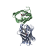
| |||||||||||||||||||||||||||||||||||||||||||||||||||||||||||||||||||||||||||||||
|---|---|---|---|---|---|---|---|---|---|---|---|---|---|---|---|---|---|---|---|---|---|---|---|---|---|---|---|---|---|---|---|---|---|---|---|---|---|---|---|---|---|---|---|---|---|---|---|---|---|---|---|---|---|---|---|---|---|---|---|---|---|---|---|---|---|---|---|---|---|---|---|---|---|---|---|---|---|---|---|---|
| 1 | 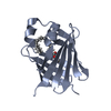
| |||||||||||||||||||||||||||||||||||||||||||||||||||||||||||||||||||||||||||||||
| 2 | 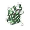
| |||||||||||||||||||||||||||||||||||||||||||||||||||||||||||||||||||||||||||||||
| Unit cell |
| |||||||||||||||||||||||||||||||||||||||||||||||||||||||||||||||||||||||||||||||
| Noncrystallographic symmetry (NCS) | NCS domain:
NCS domain segments: Ens-ID: 1
|