 Movie
Movie Controller


+ Open data

- Basic information
| Entry | Database: PDB / ID: 2fnp | ||||||
|---|---|---|---|---|---|---|---|
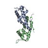
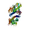
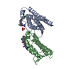
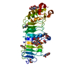
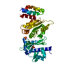
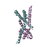
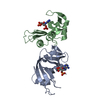
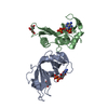
| Title | Crystal structure of SarA | ||||||
 Components Components | Staphylococcal accessory regulator A | ||||||
 Keywords Keywords | TRANSCRIPTION / wing-helix / DNA binding | ||||||
| Function / homology |  Function and homology information Function and homology informationresponse to stress / DNA-binding transcription factor activity / DNA binding / metal ion binding / cytoplasm Similarity search - Function | ||||||
| Biological species |   Staphylococcus aureus (bacteria) Staphylococcus aureus (bacteria) | ||||||
| Method |  X-RAY DIFFRACTION / SYNCHROTRON / MOLECULAR REPLACEMENT / Resolution: 2.6 Å X-RAY DIFFRACTION / SYNCHROTRON / MOLECULAR REPLACEMENT / Resolution: 2.6 Å | ||||||
 Authors Authors | Liu, Y. / Manna, A.C. / Pan, C.H. / Cheung, A.L. / Zhang, G. | ||||||
 Citation Citation | Journal: Proc.Natl.Acad.Sci.Usa / Year: 2006 Title: Structural and function analyses of the global regulatory protein SarA from Staphylococcus aureus. Authors: Liu, Y. / Manna, A.C. / Pan, C.H. / Kriksunov, I.A. / Thiel, D.J. / Cheung, A.L. / Zhang, G. | ||||||
| History |
|
- Structure visualization
| Structure viewer | Molecule: MolmilJmol/JSmol |
|---|
- Downloads & links
-Download
| PDBx/mmCIF format | 2fnp.cif.gz | 61.2 KB | Display | PDBx/mmCIF format |
|---|---|---|---|---|
| PDB format | pdb2fnp.ent.gz | 46.2 KB | Display | PDB format |
| PDBx/mmJSON format | 2fnp.json.gz | Tree view | PDBx/mmJSON format | |
| Others |  Other downloads Other downloads |
-Validation report
| Arichive directory | https://data.pdbj.org/pub/pdb/validation_reports/fn/2fnpftp://data.pdbj.org/pub/pdb/validation_reports/fn/2fnp | HTTPS FTP |
|---|
-Related structure data


-Links
 PDBj
PDBj- Assembly
| Deposited unit | 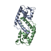
| ||||||||
|---|---|---|---|---|---|---|---|---|---|
| 1 |
| ||||||||
| Unit cell |
|
-Components
| #1: Protein | Mass: 14775.021 Da / Num. of mol.: 2 Source method: isolated from a genetically manipulated source Source: (gene. exp.) Staphylococcus aureus (bacteria) / Strain: strain MW2 / Gene: sarA / Species (production host): Escherichia coli / Production host: #2: Water | ChemComp-HOH / |  Mass: 18.015 Da / Num. of mol.: 11 / Source method: isolated from a natural source / Formula: H2O Mass: 18.015 Da / Num. of mol.: 11 / Source method: isolated from a natural source / Formula: H2O |
|---|
-Experimental details
-Experiment
| Experiment | Method: X-RAY DIFFRACTION / Number of used crystals: 1 |
|---|
- Sample preparation
| Crystal | Density Matthews: 2.8 Å3/Da / Density % sol: 55.9 % |
|---|---|
| Crystal grow | Temperature: 277 K / Method: vapor diffusion / pH: 6.5 Details: 25% PEG 8000, 0.2M calcium chloride, 0.1M cacodylate, pH 6.5, VAPOR DIFFUSION, temperature 277K |
-Data collection
| Diffraction | Mean temperature: 52.074 K |
|---|---|
| Diffraction source | Source: SYNCHROTRON / Site: ALS  / Beamline: 4.2.2 / Beamline: 4.2.2 |
| Detector | Type: ADSC QUANTUM 4 / Detector: CCD / Date: Mar 1, 2005 |
| Radiation | Protocol: SINGLE WAVELENGTH / Monochromatic (M) / Laue (L): M / Scattering type: x-ray |
| Radiation wavelength | Relative weight: 1 |
| Reflection | Resolution: 2.6→50 Å / Num. obs: 9772 / % possible obs: 96 % / Observed criterion σ(F): 0 / Observed criterion σ(I): 0 / Biso Wilson estimate: 36 Å2 / Rmerge(I) obs: 0.068 / Rsym value: 0.068 / Net I/σ(I): 12 |
| Reflection shell | Resolution: 2.6→2.69 Å / % possible all: 96 |
- Processing
| Software |
| ||||||||||||||||||||||||||||||||||||||||||||||||||||||||||||
|---|---|---|---|---|---|---|---|---|---|---|---|---|---|---|---|---|---|---|---|---|---|---|---|---|---|---|---|---|---|---|---|---|---|---|---|---|---|---|---|---|---|---|---|---|---|---|---|---|---|---|---|---|---|---|---|---|---|---|---|---|---|
| Refinement | Method to determine structure: MOLECULAR REPLACEMENT / Resolution: 2.6→39.17 Å / Rfactor Rfree error: 0.014 / Data cutoff high absF: 732953.63 / Data cutoff low absF: 0 / Isotropic thermal model: RESTRAINED / Cross valid method: THROUGHOUT / σ(F): 0
| ||||||||||||||||||||||||||||||||||||||||||||||||||||||||||||
| Solvent computation | Solvent model: FLAT MODEL / Bsol: 50.5897 Å2 / ksol: 0.325189 e/Å3 | ||||||||||||||||||||||||||||||||||||||||||||||||||||||||||||
| Displacement parameters | Biso mean: 70.5 Å2
| ||||||||||||||||||||||||||||||||||||||||||||||||||||||||||||
| Refine analyze |
| ||||||||||||||||||||||||||||||||||||||||||||||||||||||||||||
| Refinement step | Cycle: LAST / Resolution: 2.6→39.17 Å
| ||||||||||||||||||||||||||||||||||||||||||||||||||||||||||||
| Refine LS restraints |
| ||||||||||||||||||||||||||||||||||||||||||||||||||||||||||||
| LS refinement shell | Resolution: 2.6→2.76 Å / Rfactor Rfree error: 0.053 / Total num. of bins used: 6
| ||||||||||||||||||||||||||||||||||||||||||||||||||||||||||||
| Xplor file |
|
