 Movie
Movie Controller

[English] 日本語
 Yorodumi
- PDB-2f9u: HCV NS3 protease domain with NS4a peptide and a ketoamide inhibit... -
+ Open data

- Basic information
| Entry | Database: PDB / ID: 2f9u | ||||||
|---|---|---|---|---|---|---|---|
| Title | HCV NS3 protease domain with NS4a peptide and a ketoamide inhibitor with a P2 norborane | ||||||
 Components Components |
| ||||||
 Keywords Keywords | VIRAL PROTEIN / HCV / Hepatitis C protease / NS3 protease / ketoamide inhibitor | ||||||
| Function / homology |  Function and homology information Function and homology informationself proteolysis / symbiont-mediated transformation of host cell / host cell membrane / serine-type peptidase activity / host cell / symbiont entry into host cell / virion attachment to host cell / virion membrane / metal ion binding / membrane Similarity search - Function | ||||||
| Biological species |  Hepatitis C virus Hepatitis C virus | ||||||
| Method |  X-RAY DIFFRACTION / FOURIER SYNTHESIS / Resolution: 2.6 Å X-RAY DIFFRACTION / FOURIER SYNTHESIS / Resolution: 2.6 Å | ||||||
 Authors Authors | Venkatraman, S. / Njoroge, F.G. / Wu, W. / Girijavallabhan, V. / Prongay, A.J. / Butkiewicz, N. / Pichardo, J. | ||||||
 Citation Citation | Journal: Bioorg.Med.Chem.Lett. / Year: 2006 Title: Novel Inhibitors of Hepatitis C NS3-NS4A Serine Protease Derived from 2-Aza-bicyclo[2.2.1]heptane-3-carboxylic acid. Authors: Venkatraman, S. / Njoroge, F.G. / Wu, W. / Girijavallabhan, V. / Prongay, A.J. / Butkiewicz, N. / Pichardo, J. #1: Journal: Cell(Cambridge,Mass.) / Year: 1996Title: Crystal Structure of the Hepatitis C Virus NS3 Protease Domain Complexed with a Synthetic NS4a Cofactor Peptide Authors: Kim, J.L. / Morgenstern, K.A. / Lin, C. / Fox, T. / Dwyer, M.D. / Landro, J.A. / Chambers, S.P. / Markland, W. / Lepre, C.A. / O'Malley, E.T. / Harbeson, S.L. / Rice, C.M. / Murcko, M.A. / ...Authors: Kim, J.L. / Morgenstern, K.A. / Lin, C. / Fox, T. / Dwyer, M.D. / Landro, J.A. / Chambers, S.P. / Markland, W. / Lepre, C.A. / O'Malley, E.T. / Harbeson, S.L. / Rice, C.M. / Murcko, M.A. / Caron, P.R. / Thomson, J.A. #2: Journal: Bioorg.Med.Chem.Lett. / Year: 2005Title: Hepatitis C virus NS3-4A serine protease inhibitors: Use of a P2-P1 cyclopropyl alanine combination to improve potency. Authors: Bogen, S. / Saksena, A.K. / Arasappan, A. / Gu, N. / Njoroge, F.G. / Girijavallabhan, V. / Pichardo, J. / Butkiewicz, N. / Prongay, A. / Madison, A. #3: Journal: J.Med.Chem. / Year: 2005 Title: Design and Synthesis of Depeptidized Macrocyclic Inhibitors of Hepatitis C NS3-4A Protease Using Structure-Based Drug Design Authors: Venkatraman, S. / Njoroge, F.G. / Girijavallabhan, V.M. / Madison, V.S. / Yao, N.H. / Prongay, A.J. / Butkiewicz, N. / Pichardo, J. #4: Journal: Angew.Chem.Int.Ed.Engl. / Year: 2005 Title: Proline-Based Macrocyclic Inhibitors of the Hepatitis C Virus: Stereoselective Synthesis and Biological Activity. Authors: Chen, K.X. / Njoroge, F.G. / Vibulbhan, B. / Prongay, A. / Pichardo, J. / Madison, V. / Buevich, A. / Chan, T.M. #5: Journal: Bioorg.Med.Chem.Lett. / Year: 2004 Title: Novel 2-oxoimidazolidine-4-carboxylic acid derivatives as Hepatitis C virus NS3-4A serine protease inhibitors: synthesis, activity and X-ray crystal structure of an enzyme inhibitor complex Authors: Arasappan, A. / Njoroge, F.G. / Parekh, T.N. / Yang, X. / Pichardo, J. / Butkiewicz, N. / Prongay, A. / Yao, N. / Girijavallabhan, V. | ||||||
| History |
|
- Structure visualization
| Structure viewer | Molecule: MolmilJmol/JSmol |
|---|
- Downloads & links
-Download
| PDBx/mmCIF format | 2f9u.cif.gz | 90.3 KB | Display | PDBx/mmCIF format |
|---|---|---|---|---|
| PDB format | pdb2f9u.ent.gz | 67.5 KB | Display | PDB format |
| PDBx/mmJSON format | 2f9u.json.gz | Tree view | PDBx/mmJSON format | |
| Others |  Other downloads Other downloads |
-Validation report
| Arichive directory | https://data.pdbj.org/pub/pdb/validation_reports/f9/2f9uftp://data.pdbj.org/pub/pdb/validation_reports/f9/2f9u | HTTPS FTP |
|---|
-Related structure data
| Related structure data | |
|---|---|
| Similar structure data |


-Links
 PDBj
PDBj
- Assembly
| Deposited unit | 
| ||||||||
|---|---|---|---|---|---|---|---|---|---|
| 1 |
| ||||||||
| Unit cell |
| ||||||||
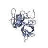
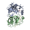
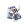
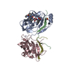
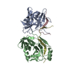
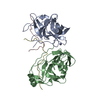
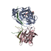
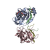
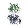
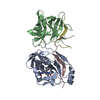
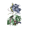
| Details | The NS3 protease domain, residues 1-181 of NS3, exists in a complex with an NS4a peptide and an inhibitor. There is a dimer of the NS3 domain-NS4a peptide complex, but only one monomer (Chains A and B) have the inhibitor bound to the active site. This dimer is the component of the asymmetric unit. In vivo, the protease domain is part of a multi enzyme protein (having both protease and helicase activities). The NS3 protease domain with the NS4A peptide is known to be catalytically active in the absence of the helicase domain, although it is not known whether it is active as a monomer or dimer. |
-Components
| #1: Protein | Mass: 21102.027 Da / Num. of mol.: 2 / Fragment: protease domain (Residues : 1-181) Source method: isolated from a genetically manipulated source Source: (gene. exp.) Hepatitis C virus / Genus: Hepacivirus / Strain: H StrainGene: NS3 protease domain ( residues 1027-1207 of the polyprotein). Plasmid: NS3(181)His6/pET22b / Production host:  #2: Protein/peptide | Mass: 2394.039 Da / Num. of mol.: 2 / Fragment: Residues: 21-39 / Source method: obtained synthetically Details: Solid-phase peptide synthesis of the NS4a residues 21-39 peptide with N-terminal KK and C-terminal KK extensions. References: GenBank: 51039195 #3: Chemical |   Mass: 65.409 Da / Num. of mol.: 2 / Source method: obtained synthetically / Formula: Zn / Details: ketoamide peptide Mass: 65.409 Da / Num. of mol.: 2 / Source method: obtained synthetically / Formula: Zn / Details: ketoamide peptide#4: Chemical | ChemComp-5NH / | 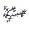  Mass: 738.913 Da / Num. of mol.: 1 / Source method: obtained synthetically / Formula: C39H58N6O8 Mass: 738.913 Da / Num. of mol.: 1 / Source method: obtained synthetically / Formula: C39H58N6O8#5: Water | ChemComp-HOH / |  Mass: 18.015 Da / Num. of mol.: 115 / Source method: isolated from a natural source / Formula: H2O Mass: 18.015 Da / Num. of mol.: 115 / Source method: isolated from a natural source / Formula: H2OHas protein modification | Y | |
|---|
-Experimental details
-Experiment
| Experiment | Method: X-RAY DIFFRACTION / Number of used crystals: 1 |
|---|
- Sample preparation
| Crystal | Density Matthews: 3.91 Å3/Da / Density % sol: 68.54 % |
|---|---|
| Crystal grow | Temperature: 285 K / pH: 5.7 Details: 12-15 mG/mL Protein in 15 mM MES, pH 6.5 - 1.0 M NaCl equivolume mixture with (0.85-1.05M) NaCl- 0.1M MES-0.1 M Na/KPO4, pH5.5-5.8 - 5mM BME, equilibrated with 1.35-1.55M NaCl-0.1M MES-0.1M ...Details: 12-15 mG/mL Protein in 15 mM MES, pH 6.5 - 1.0 M NaCl equivolume mixture with (0.85-1.05M) NaCl- 0.1M MES-0.1 M Na/KPO4, pH5.5-5.8 - 5mM BME, equilibrated with 1.35-1.55M NaCl-0.1M MES-0.1M Na/KPO4, pH 5.6-5.8 - 5 mM BME, VAPOR DIFFUSION, HANGING DROP, temperature 285K, pH 5.70 |
-Data collection
| Diffraction | Mean temperature: 95 K |
|---|---|
| Diffraction source | Source: ROTATING ANODE / Type: RIGAKU RUH3R / Wavelength: 1.5418 |
| Detector | Type: RIGAKU RAXIS IV / Detector: IMAGE PLATE / Date: Oct 25, 2005 / Details: OSMIC GREEN |
| Radiation | Monochromator: OSMIC GREEN / Protocol: SINGLE WAVELENGTH / Monochromatic (M) / Laue (L): M / Scattering type: x-ray |
| Radiation wavelength | Wavelength: 1.5418 Å / Relative weight: 1 |
| Reflection | Resolution: 2.6→50 Å / Num. obs: 20873 / % possible obs: 92.7 % / Observed criterion σ(I): 1.3 / Redundancy: 4.6 % / Biso Wilson estimate: 35.89 Å2 / Rmerge(I) obs: 0.075 / Net I/σ(I): 18.4 |
| Reflection shell | Resolution: 2.6→2.66 Å / Redundancy: 1.6 % / Rmerge(I) obs: 0.49 / Mean I/σ(I) obs: 1.3 / % possible all: 42 |
- Processing
| Software |
| ||||||||||||||||||||||||||||||||||||||||||||||||||||||||||||
|---|---|---|---|---|---|---|---|---|---|---|---|---|---|---|---|---|---|---|---|---|---|---|---|---|---|---|---|---|---|---|---|---|---|---|---|---|---|---|---|---|---|---|---|---|---|---|---|---|---|---|---|---|---|---|---|---|---|---|---|---|---|
| Refinement | Method to determine structure: FOURIER SYNTHESIS / Resolution: 2.6→8 Å / σ(F): 1 / Stereochemistry target values: Engh & Huber
| ||||||||||||||||||||||||||||||||||||||||||||||||||||||||||||
| Refinement step | Cycle: LAST / Resolution: 2.6→8 Å
| ||||||||||||||||||||||||||||||||||||||||||||||||||||||||||||
| Refine LS restraints |
| ||||||||||||||||||||||||||||||||||||||||||||||||||||||||||||
| LS refinement shell | Resolution: 2.6→2.71 Å / Total num. of bins used: 8
|
