 Movie
Movie Controller

[English] 日本語
 Yorodumi
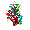
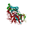
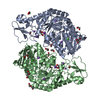
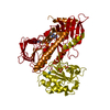
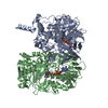
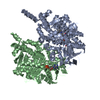
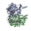
- PDB-2e1m: Crystal Structure of L-Glutamate Oxidase from Streptomyces sp. X-119-6 -
+ Open data

- Basic information
| Entry | Database: PDB / ID: 2e1m | ||||||
|---|---|---|---|---|---|---|---|
| Title | Crystal Structure of L-Glutamate Oxidase from Streptomyces sp. X-119-6 | ||||||
 Components Components | (L-glutamate oxidase) x 3 | ||||||
 Keywords Keywords | OXIDOREDUCTASE / L-amino acid oxidase / L-Glutamate Oxidase / FAD / L-GOX / Flavoprotein | ||||||
| Function / homology |  Function and homology information Function and homology informationL-glutamate oxidase / L-amino-acid oxidase activity / amino acid catabolic process / nucleotide binding / extracellular region Similarity search - Function | ||||||
| Biological species |  Streptomyces sp. (bacteria) Streptomyces sp. (bacteria) | ||||||
| Method |  X-RAY DIFFRACTION / SYNCHROTRON / MOLECULAR REPLACEMENT / Resolution: 2.8 Å X-RAY DIFFRACTION / SYNCHROTRON / MOLECULAR REPLACEMENT / Resolution: 2.8 Å | ||||||
 Authors Authors | Sasaki, C. / Kashima, A. / Sakaguchi, C. / Mizuno, H. / Arima, J. / Kusakabe, H. / Tamura, T. / Sugio, S. / Inagaki, K. | ||||||
 Citation Citation | Journal: Febs J. / Year: 2009 Title: Structural characterization of l-glutamate oxidase from Streptomyces sp. X-119-6 Authors: Arima, J. / Sasaki, C. / Sakaguchi, C. / Mizuno, H. / Tamura, T. / Kashima, A. / Kusakabe, H. / Sugio, S. / Inagaki, K. | ||||||
| History |
|
- Structure visualization
| Structure viewer | Molecule: MolmilJmol/JSmol |
|---|
- Downloads & links
-Download
| PDBx/mmCIF format | 2e1m.cif.gz | 136.9 KB | Display | PDBx/mmCIF format |
|---|---|---|---|---|
| PDB format | pdb2e1m.ent.gz | 103.6 KB | Display | PDB format |
| PDBx/mmJSON format | 2e1m.json.gz | Tree view | PDBx/mmJSON format | |
| Others |  Other downloads Other downloads |
-Validation report
| Summary document | 2e1m_validation.pdf.gz | 739.4 KB | Display | wwPDB validaton report |
|---|---|---|---|---|
| Full document | 2e1m_full_validation.pdf.gz | 763.4 KB | Display | |
| Data in XML | 2e1m_validation.xml.gz | 25.8 KB | Display | |
| Data in CIF | 2e1m_validation.cif.gz | 34.1 KB | Display | |
| Arichive directory | https://data.pdbj.org/pub/pdb/validation_reports/e1/2e1mftp://data.pdbj.org/pub/pdb/validation_reports/e1/2e1m | HTTPS FTP |
-Related structure data
| Related structure data | 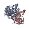 1f8rS S: Starting model for refinement |
|---|---|
| Similar structure data |


-Links
 PDBj
PDBj
- Assembly
| Deposited unit | 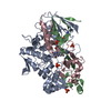
| ||||||||
|---|---|---|---|---|---|---|---|---|---|
| 1 | 
| ||||||||
| Unit cell |
|
-Components
| #1: Protein | Mass: 42019.227 Da / Num. of mol.: 1 / Fragment: N-terminal domain, residues 15-390 Source method: isolated from a genetically manipulated source Source: (gene. exp.) Streptomyces sp. (bacteria) / Strain: X-119-6 / Plasmid: pKK223-3 / Production host: | ||||
|---|---|---|---|---|---|
| #2: Protein | Mass: 15057.732 Da / Num. of mol.: 1 / Fragment: residues 391-520 Source method: isolated from a genetically manipulated source Source: (gene. exp.) Streptomyces sp. (bacteria) / Strain: X-119-6 / Plasmid: pKK223-3 / Production host: | ||||
| #3: Protein | Mass: 19354.182 Da / Num. of mol.: 1 / Fragment: C-terminal domain, residues 521-701 Source method: isolated from a genetically manipulated source Source: (gene. exp.) Streptomyces sp. (bacteria) / Strain: X-119-6 / Plasmid: pKK223-3 / Production host: | ||||
| #4: Chemical |   Mass: 94.971 Da / Num. of mol.: 3 / Source method: obtained synthetically / Formula: PO4 Mass: 94.971 Da / Num. of mol.: 3 / Source method: obtained synthetically / Formula: PO4#5: Chemical | ChemComp-FAD / |   Mass: 785.550 Da / Num. of mol.: 1 / Source method: obtained synthetically / Formula: C27H33N9O15P2 / Comment: FAD*YM Mass: 785.550 Da / Num. of mol.: 1 / Source method: obtained synthetically / Formula: C27H33N9O15P2 / Comment: FAD*YMSequence details | THE PRECURSOR OF L-GLUTAMETE OXIDASE IS EXPRESSED AS ONE POLYPEPTIDE CHAIN AND MATURED BY ...THE PRECURSOR OF L-GLUTAMETE OXIDASE IS EXPRESSED AS ONE POLYPEPTID | |
-Experimental details
-Experiment
| Experiment | Method: X-RAY DIFFRACTION / Number of used crystals: 1 |
|---|
- Sample preparation
| Crystal | Density Matthews: 2.39 Å3/Da / Density % sol: 48.62 % |
|---|---|
| Crystal grow | Temperature: 278 K / Method: vapor diffusion, sitting drop / pH: 10.5 Details: 100mM CAPS-buffer, 1200mM NaH2PO4, 800mM K2HPO4, 50mM LiSO4, 5mM DTT, 20mM KPB, 10mg/ml GOX, pH 10.50, VAPOR DIFFUSION, SITTING DROP, temperature 278K |
-Data collection
| Diffraction | Mean temperature: 100 K |
|---|---|
| Diffraction source | Source: SYNCHROTRON / Site: SPring-8  / Beamline: BL24XU / Wavelength: 1 / Beamline: BL24XU / Wavelength: 1 |
| Detector | Type: RIGAKU RAXIS V / Detector: IMAGE PLATE / Date: Nov 22, 2004 |
| Radiation | Monochromator: DIAMOND / Protocol: SINGLE WAVELENGTH / Monochromatic (M) / Laue (L): M / Scattering type: x-ray |
| Radiation wavelength | Wavelength: 1 Å / Relative weight: 1 |
| Reflection | Resolution: 2.6→50 Å / Num. obs: 24388 / % possible obs: 100 % / Redundancy: 10 % / Biso Wilson estimate: 75.5 Å2 / Rmerge(I) obs: 0.08 / Net I/σ(I): 34.043 |
| Reflection shell | Resolution: 2.6→2.69 Å / Redundancy: 9.8 % / Rmerge(I) obs: 0.489 / Mean I/σ(I) obs: 4.779 / % possible all: 100 |
- Processing
| Software |
| ||||||||||||||||||||||||||||||||||||||||||||||||||||||||||||
|---|---|---|---|---|---|---|---|---|---|---|---|---|---|---|---|---|---|---|---|---|---|---|---|---|---|---|---|---|---|---|---|---|---|---|---|---|---|---|---|---|---|---|---|---|---|---|---|---|---|---|---|---|---|---|---|---|---|---|---|---|---|
| Refinement | Method to determine structure: MOLECULAR REPLACEMENT Starting model: PDB ENTRY 1F8R Resolution: 2.8→45.27 Å / Rfactor Rfree error: 0.01 / Data cutoff high absF: 2982255.12 / Data cutoff low absF: 0 / Cross valid method: THROUGHOUT / σ(F): 0 / Stereochemistry target values: ENGH & HUBER
| ||||||||||||||||||||||||||||||||||||||||||||||||||||||||||||
| Displacement parameters | Biso mean: 34.3 Å2
| ||||||||||||||||||||||||||||||||||||||||||||||||||||||||||||
| Refine analyze |
| ||||||||||||||||||||||||||||||||||||||||||||||||||||||||||||
| Refinement step | Cycle: LAST / Resolution: 2.8→45.27 Å
| ||||||||||||||||||||||||||||||||||||||||||||||||||||||||||||
| Refine LS restraints |
| ||||||||||||||||||||||||||||||||||||||||||||||||||||||||||||
| LS refinement shell | Resolution: 2.8→2.85 Å / Rfactor Rfree error: 0.039 / Total num. of bins used: 6
|
