 Movie
Movie Controller

[English] 日本語
 Yorodumi
- PDB-1f8r: CRYSTAL STRUCTURE OF L-AMINO ACID OXIDASE FROM CALLOSELASMA RHODO... -
+ Open data

- Basic information
| Entry | Database: PDB / ID: 1f8r | |||||||||
|---|---|---|---|---|---|---|---|---|---|---|
| Title | CRYSTAL STRUCTURE OF L-AMINO ACID OXIDASE FROM CALLOSELASMA RHODOSTOMA COMPLEXED WITH CITRATE | |||||||||
 Components Components | L-AMINO ACID OXIDASE | |||||||||
 Keywords Keywords | OXIDOREDUCTASE / FLAVOENZYME / OXIDASE / ENANTIOMERIC SPECIFICITY / ACTIVE SITE FUNNEL / HELICAL DOMAIN / FAD-BINDING DOMAIN | |||||||||
| Function / homology |  Function and homology information Function and homology informationL-phenylalaine oxidase activity / L-amino-acid oxidase / amino acid catabolic process / toxin activity / killing of cells of another organism / defense response to bacterium / apoptotic process / extracellular region Similarity search - Function | |||||||||
| Biological species |  Calloselasma rhodostoma (Malayan pit viper) Calloselasma rhodostoma (Malayan pit viper) | |||||||||
| Method |  X-RAY DIFFRACTION / SYNCHROTRON / Resolution: 2 Å X-RAY DIFFRACTION / SYNCHROTRON / Resolution: 2 Å | |||||||||
 Authors Authors | Pawelek, P.D. / Cheah, J. / Coulombe, R. / Macheroux, P. / Ghisla, S. / Vrielink, A. | |||||||||
 Citation Citation | Journal: EMBO J. / Year: 2000 Title: The structure of L-amino acid oxidase reveals the substrate trajectory into an enantiomerically conserved active site. Authors: Pawelek, P.D. / Cheah, J. / Coulombe, R. / Macheroux, P. / Ghisla, S. / Vrielink, A. | |||||||||
| History |
|
- Structure visualization
| Structure viewer | Molecule: MolmilJmol/JSmol |
|---|
- Downloads & links
-Download
| PDBx/mmCIF format | 1f8r.cif.gz | 439.5 KB | Display | PDBx/mmCIF format |
|---|---|---|---|---|
| PDB format | pdb1f8r.ent.gz | 355.5 KB | Display | PDB format |
| PDBx/mmJSON format | 1f8r.json.gz | Tree view | PDBx/mmJSON format | |
| Others |  Other downloads Other downloads |
-Validation report
| Arichive directory | https://data.pdbj.org/pub/pdb/validation_reports/f8/1f8rftp://data.pdbj.org/pub/pdb/validation_reports/f8/1f8r | HTTPS FTP |
|---|
-Related structure data


-Links
 PDBj
PDBj
- Assembly
| Deposited unit | 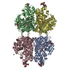
| |||||||||
|---|---|---|---|---|---|---|---|---|---|---|
| 1 | 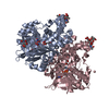
| |||||||||
| 2 | 
| |||||||||
| Unit cell |
| |||||||||
| Noncrystallographic symmetry (NCS) | NCS domain:
| |||||||||
| Details | Biological unit is a dimer. |
-Components
-Protein , 1 types, 4 molecules ABCD
| #1: Protein | Mass: 56299.258 Da / Num. of mol.: 4 / Source method: isolated from a natural source Source: (natural) Calloselasma rhodostoma (Malayan pit viper)Secretion: VENOM References: GenBank: 6850960, UniProt: P81382*PLUS, L-amino-acid oxidase |
|---|
-Sugars , 2 types, 8 molecules 
| #2: Polysaccharide | 2-acetamido-2-deoxy-beta-D-glucopyranose-(1-4)-[alpha-L-fucopyranose-(1-6)]2-acetamido-2-deoxy-beta- ...2-acetamido-2-deoxy-beta-D-glucopyranose-(1-4)-[alpha-L-fucopyranose-(1-6)]2-acetamido-2-deoxy-beta-D-glucopyranose Source method: isolated from a genetically manipulated source #3: Sugar | ChemComp-NAG /  Type: D-saccharide, beta linking / Mass: 221.208 Da / Num. of mol.: 4 Type: D-saccharide, beta linking / Mass: 221.208 Da / Num. of mol.: 4Source method: isolated from a genetically manipulated source Formula: C8H15NO6 |
|---|
-Non-polymers , 3 types, 1877 molecules 


| #4: Chemical | ChemComp-CIT /  Mass: 192.124 Da / Num. of mol.: 4 / Source method: obtained synthetically / Formula: C6H8O7 Mass: 192.124 Da / Num. of mol.: 4 / Source method: obtained synthetically / Formula: C6H8O7#5: Chemical | ChemComp-FAD /  Mass: 785.550 Da / Num. of mol.: 4 / Source method: obtained synthetically / Formula: C27H33N9O15P2 / Comment: FAD*YM Mass: 785.550 Da / Num. of mol.: 4 / Source method: obtained synthetically / Formula: C27H33N9O15P2 / Comment: FAD*YM#6: Water | ChemComp-HOH / | Mass: 18.015 Da / Num. of mol.: 1869 / Source method: isolated from a natural source / Formula: H2O |
|---|
-Details
| Has protein modification | Y |
|---|---|
| Sequence details | F0-FC DENSITY OBSERVED EXTENDING FROM ALA149 SUGGESTING |
-Experimental details
-Experiment
| Experiment | Method: X-RAY DIFFRACTION / Number of used crystals: 1 |
|---|
- Sample preparation
| Crystal | Density Matthews: 2.65 Å3/Da / Density % sol: 53.64 % |
|---|---|
| Crystal grow | Temperature: 290 K / Method: vapor diffusion, hanging drop / pH: 4.5 Details: PEG4000, ammonium sulfate, sodium citrate, glycerol, pH 4.5, VAPOR DIFFUSION, HANGING DROP, temperature 290.0K |
| Crystal grow | *PLUS Details: Jancarik, J., (1991) J. Appl. Crystallogr., 24, 409. |
| Components of the solutions | *PLUS Conc.: 10 mg/ml / Common name: protein |
-Data collection
| Diffraction | Mean temperature: 83 K |
|---|---|
| Diffraction source | Source: SYNCHROTRON / Site: NSLS  / Beamline: X8C / Wavelength: 1.072 / Beamline: X8C / Wavelength: 1.072 |
| Detector | Type: MARRESEARCH / Detector: AREA DETECTOR / Date: Aug 16, 1998 |
| Radiation | Protocol: SINGLE WAVELENGTH / Monochromatic (M) / Laue (L): M / Scattering type: x-ray |
| Radiation wavelength | Wavelength: 1.072 Å / Relative weight: 1 |
| Reflection | Resolution: 2→50 Å / Num. all: 158011 / Num. obs: 139242 / % possible obs: 88.1 % / Observed criterion σ(F): 0 / Observed criterion σ(I): 0 / Redundancy: 2.8 % / Biso Wilson estimate: 13.6 Å2 / Rmerge(I) obs: 0.046 / Net I/σ(I): 18.9 |
| Reflection shell | Resolution: 2→2.07 Å / Redundancy: 2.01 % / Rmerge(I) obs: 0.159 / Num. unique all: 11263 / % possible all: 71.7 |
| Reflection | *PLUS Num. measured all: 391000 |
- Processing
| Software |
| ||||||||||||||||||||||||||||||||||||||||
|---|---|---|---|---|---|---|---|---|---|---|---|---|---|---|---|---|---|---|---|---|---|---|---|---|---|---|---|---|---|---|---|---|---|---|---|---|---|---|---|---|---|
| Refinement | Resolution: 2→500 Å / Rfactor Rfree error: 0.002 / Data cutoff high absF: 2553955.22 / Data cutoff low absF: 0 / Isotropic thermal model: RESTRAINED / Cross valid method: THROUGHOUT / σ(F): 0 / σ(I): 0 / Stereochemistry target values: ENGH & HUBER
| ||||||||||||||||||||||||||||||||||||||||
| Solvent computation | Solvent model: FLAT MODEL / Bsol: 57.6 Å2 / ksol: 0.388 e/Å3 | ||||||||||||||||||||||||||||||||||||||||
| Displacement parameters | Biso mean: 24.5 Å2
| ||||||||||||||||||||||||||||||||||||||||
| Refine analyze |
| ||||||||||||||||||||||||||||||||||||||||
| Refinement step | Cycle: LAST / Resolution: 2→500 Å
| ||||||||||||||||||||||||||||||||||||||||
| Refine LS restraints |
| ||||||||||||||||||||||||||||||||||||||||
| Refine LS restraints NCS |
| ||||||||||||||||||||||||||||||||||||||||
| LS refinement shell | Resolution: 2→2.13 Å / Rfactor Rfree error: 0.006 / Total num. of bins used: 6
| ||||||||||||||||||||||||||||||||||||||||
| Xplor file |
| ||||||||||||||||||||||||||||||||||||||||
| Software | *PLUS Name: CNS / Version: 0.5 / Classification: refinement | ||||||||||||||||||||||||||||||||||||||||
| Refinement | *PLUS Lowest resolution: 50 Å / σ(F): 0 / % reflection Rfree: 10 % / Rfactor obs: 0.185 / Rfactor Rfree: 0.21 | ||||||||||||||||||||||||||||||||||||||||
| Solvent computation | *PLUS | ||||||||||||||||||||||||||||||||||||||||
| Displacement parameters | *PLUS Biso mean: 24.5 Å2 | ||||||||||||||||||||||||||||||||||||||||
| Refine LS restraints | *PLUS
| ||||||||||||||||||||||||||||||||||||||||
| LS refinement shell | *PLUS Rfactor Rfree: 0.248 / % reflection Rfree: 10 % / Rfactor Rwork: 0.214 |
