 Movie
Movie Controller


+ Open data

- Basic information
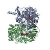
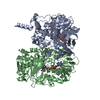
| Entry | Database: PDB / ID: 7e0c | ||||||
|---|---|---|---|---|---|---|---|
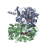
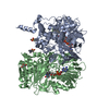
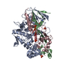
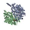
| Title | Structure of L-glutamate oxidase R305E mutant | ||||||
 Components Components | L-glutamate oxidase | ||||||
 Keywords Keywords | OXIDOREDUCTASE / L-glutamic acid oxidase / L-amino acid oxidase | ||||||
| Function / homology |  Function and homology information Function and homology informationL-glutamate oxidase / L-glutamate oxidase activity / amino acid catabolic process / nucleotide binding / extracellular region Similarity search - Function | ||||||
| Biological species |  Streptomyces sp. X-119-6 (bacteria) Streptomyces sp. X-119-6 (bacteria) | ||||||
| Method |  X-RAY DIFFRACTION / SYNCHROTRON / MOLECULAR REPLACEMENT / Resolution: 2.65 Å X-RAY DIFFRACTION / SYNCHROTRON / MOLECULAR REPLACEMENT / Resolution: 2.65 Å | ||||||
 Authors Authors | Ito, N. / Matsuo, S. / Inagaki, K. / Imada, K. | ||||||
| Funding support |  Japan, 1items Japan, 1items
| ||||||
 Citation Citation | Journal: Protein Sci. / Year: 2021 Title: A new l-arginine oxidase engineered from l-glutamate oxidase. Authors: Yano, Y. / Matsuo, S. / Ito, N. / Tamura, T. / Kusakabe, H. / Inagaki, K. / Imada, K. | ||||||
| History |
|
- Structure visualization
| Structure viewer | Molecule: MolmilJmol/JSmol |
|---|
- Downloads & links
-Download
| PDBx/mmCIF format | 7e0c.cif.gz | 139.5 KB | Display | PDBx/mmCIF format |
|---|---|---|---|---|
| PDB format | pdb7e0c.ent.gz | 104.2 KB | Display | PDB format |
| PDBx/mmJSON format | 7e0c.json.gz | Tree view | PDBx/mmJSON format | |
| Others |  Other downloads Other downloads |
-Validation report
| Arichive directory | https://data.pdbj.org/pub/pdb/validation_reports/e0/7e0cftp://data.pdbj.org/pub/pdb/validation_reports/e0/7e0c | HTTPS FTP |
|---|
-Related structure data
| Related structure data |  7e0dC  2e1mS S: Starting model for refinement C: citing same article ( |
|---|---|
| Similar structure data |

-Links
 PDBj
PDBj
- Assembly
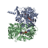
| Deposited unit | 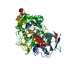
| ||||||||
|---|---|---|---|---|---|---|---|---|---|
| 1 | 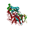
| ||||||||
| Unit cell |
| ||||||||
| Components on special symmetry positions |
|
-Components
| #1: Protein | Mass: 76427.250 Da / Num. of mol.: 1 / Mutation: R305E Source method: isolated from a genetically manipulated source Source: (gene. exp.) Streptomyces sp. X-119-6 (bacteria) / Gene: Lgox / Plasmid: pMal-c2 / Production host: |
|---|---|
| #2: Chemical | ChemComp-FAD /   Mass: 785.550 Da / Num. of mol.: 1 / Source method: obtained synthetically / Formula: C27H33N9O15P2 / Feature type: SUBJECT OF INVESTIGATION / Comment: FAD*YM Mass: 785.550 Da / Num. of mol.: 1 / Source method: obtained synthetically / Formula: C27H33N9O15P2 / Feature type: SUBJECT OF INVESTIGATION / Comment: FAD*YM |
| #3: Water | ChemComp-HOH /  Mass: 18.015 Da / Num. of mol.: 87 / Source method: isolated from a natural source / Formula: H2O Mass: 18.015 Da / Num. of mol.: 87 / Source method: isolated from a natural source / Formula: H2O |
| Has ligand of interest | Y |
-Experimental details
-Experiment
| Experiment | Method: X-RAY DIFFRACTION / Number of used crystals: 1 |
|---|
- Sample preparation
| Crystal | Density Matthews: 2.45 Å3/Da / Density % sol: 49.74 % |
|---|---|
| Crystal grow | Temperature: 277 K / Method: vapor diffusion, sitting drop / pH: 7 Details: 12% (w/v) PEG 8000, 0.1M Tris-HCl pH7.0, 75mM MgCl2 |
-Data collection
| Diffraction | Mean temperature: 100 K / Serial crystal experiment: N |
|---|---|
| Diffraction source | Source: SYNCHROTRON / Site: SPring-8 / Beamline: BL41XU / Wavelength: 1 Å |
| Detector | Type: DECTRIS PILATUS 6M / Detector: PIXEL / Date: Oct 8, 2016 |
| Radiation | Monochromator: Double-crystal monochromator / Protocol: SINGLE WAVELENGTH / Monochromatic (M) / Laue (L): M / Scattering type: x-ray |
| Radiation wavelength | Wavelength: 1 Å / Relative weight: 1 |
| Reflection | Resolution: 2.65→84.3 Å / Num. obs: 22897 / % possible obs: 99.9 % / Redundancy: 12.3 % / Biso Wilson estimate: 36.5 Å2 / CC1/2: 0.999 / Rmerge(I) obs: 0.115 / Rpim(I) all: 0.034 / Net I/σ(I): 18.5 |
| Reflection shell | Resolution: 2.65→2.78 Å / Redundancy: 13 % / Rmerge(I) obs: 0.67 / Mean I/σ(I) obs: 4.6 / Num. unique obs: 2970 / CC1/2: 0.93 / Rpim(I) all: 0.191 / % possible all: 100 |
- Processing
| Software |
| |||||||||||||||||||||||||||||||||||||||||||||||||||||||||||||||
|---|---|---|---|---|---|---|---|---|---|---|---|---|---|---|---|---|---|---|---|---|---|---|---|---|---|---|---|---|---|---|---|---|---|---|---|---|---|---|---|---|---|---|---|---|---|---|---|---|---|---|---|---|---|---|---|---|---|---|---|---|---|---|---|---|
| Refinement | Method to determine structure: MOLECULAR REPLACEMENT Starting model: 2E1M Resolution: 2.65→66.311 Å / SU ML: 0.26 / Cross valid method: FREE R-VALUE / σ(F): 1.35 / Phase error: 22.69 / Stereochemistry target values: ML
| |||||||||||||||||||||||||||||||||||||||||||||||||||||||||||||||
| Solvent computation | Shrinkage radii: 0.9 Å / VDW probe radii: 1.11 Å / Solvent model: FLAT BULK SOLVENT MODEL | |||||||||||||||||||||||||||||||||||||||||||||||||||||||||||||||
| Refinement step | Cycle: LAST / Resolution: 2.65→66.311 Å
| |||||||||||||||||||||||||||||||||||||||||||||||||||||||||||||||
| Refine LS restraints |
| |||||||||||||||||||||||||||||||||||||||||||||||||||||||||||||||
| LS refinement shell |
|
