 Movie
Movie Controller


+ Open data

- Basic information
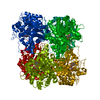
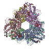
| Entry | Database: PDB / ID: 2dga | ||||||
|---|---|---|---|---|---|---|---|
| Title | Crystal structure of hexameric beta-glucosidase in wheat | ||||||
 Components Components | Beta-glucosidase | ||||||
 Keywords Keywords | HYDROLASE / alpha/beta barrel | ||||||
| Function / homology |  Function and homology information4-hydroxy-7-methoxy-3-oxo-3,4-dihydro-2H-1,4-benzoxazin-2-yl glucoside beta-D-glucosidase / DIMBOA glucoside beta-D-glucosidase activity / scopolin beta-glucosidase activity / beta-glucosidase activity / beta-glucosidase / chloroplast / carbohydrate metabolic process Function and homology information4-hydroxy-7-methoxy-3-oxo-3,4-dihydro-2H-1,4-benzoxazin-2-yl glucoside beta-D-glucosidase / DIMBOA glucoside beta-D-glucosidase activity / scopolin beta-glucosidase activity / beta-glucosidase activity / beta-glucosidase / chloroplast / carbohydrate metabolic processSimilarity search - Function | ||||||
| Biological species |  Triticum aestivum (bread wheat) Triticum aestivum (bread wheat) | ||||||
| Method | X-RAY DIFFRACTION / SYNCHROTRON / MOLECULAR REPLACEMENT / Resolution: 1.8 Å | ||||||
 Authors Authors | Sue, M. / Yamazaki, K. / Miyamoto, T. / Yajima, S. | ||||||
 Citation Citation | Journal: Plant Physiol. / Year: 2006 Title: Molecular and Structural Characterization of Hexameric beta-D-Glucosidases in Wheat and Rye. Authors: Sue, M. / Yamazaki, K. / Yajima, S. / Nomura, T. / Matsukawa, T. / Iwamura, H. / Miyamoto, T. | ||||||
| History |
|
- Structure visualization
| Structure viewer | Molecule: MolmilJmol/JSmol |
|---|
- Downloads & links
-Download
| PDBx/mmCIF format | 2dga.cif.gz | 129.2 KB | Display | PDBx/mmCIF format |
|---|---|---|---|---|
| PDB format | pdb2dga.ent.gz | 96 KB | Display | PDB format |
| PDBx/mmJSON format | 2dga.json.gz | Tree view | PDBx/mmJSON format | |
| Others |  Other downloads Other downloads |
-Validation report
| Arichive directory | https://data.pdbj.org/pub/pdb/validation_reports/dg/2dgaftp://data.pdbj.org/pub/pdb/validation_reports/dg/2dga | HTTPS FTP |
|---|
-Related structure data
| Related structure data |  1v03S S: Starting model for refinement |
|---|---|
| Similar structure data |

-Links
 PDBj
PDBj- Assembly
| Deposited unit | 
| ||||||||
|---|---|---|---|---|---|---|---|---|---|
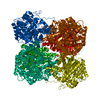
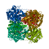
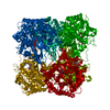
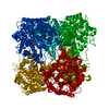
| 1 | x 6
| ||||||||
| Unit cell |
|
-Components
| #1: Protein | / beta-D-glucosidase Mass: 64166.574 Da / Num. of mol.: 1 / Fragment: residues 1-520 Source method: isolated from a genetically manipulated source Source: (gene. exp.) Triticum aestivum (bread wheat) / Gene: Taglu1b / Plasmid: pET30a / Production host:  Escherichia coli (E. coli) / Strain (production host): BL21-CodonPlus(DE3)-RIL / References: UniProt: Q1XH05, beta-glucosidase Escherichia coli (E. coli) / Strain (production host): BL21-CodonPlus(DE3)-RIL / References: UniProt: Q1XH05, beta-glucosidase | ||
|---|---|---|---|
| #2: Chemical | ChemComp-SO4 / Sulfate  Mass: 96.063 Da / Num. of mol.: 1 / Source method: obtained synthetically / Formula: SO4 Mass: 96.063 Da / Num. of mol.: 1 / Source method: obtained synthetically / Formula: SO4 | ||
| #3: Chemical | Glycerol  Mass: 92.094 Da / Num. of mol.: 3 / Source method: obtained synthetically / Formula: C3H8O3 Mass: 92.094 Da / Num. of mol.: 3 / Source method: obtained synthetically / Formula: C3H8O3#4: Water | ChemComp-HOH / | Water Mass: 18.015 Da / Num. of mol.: 613 / Source method: isolated from a natural source / Formula: H2O Mass: 18.015 Da / Num. of mol.: 613 / Source method: isolated from a natural source / Formula: H2O |
-Experimental details
-Experiment
| Experiment | Method: X-RAY DIFFRACTION / Number of used crystals: 1 |
|---|
- Sample preparation
| Crystal | Density Matthews: 4.79 Å3/Da / Density % sol: 74.3 % / Description: the file contains Friedel pairs. |
|---|---|
| Crystal grow | Temperature: 293 K / Method: vapor diffusion, hanging drop / pH: 7.2 Details: 10mM HEPES pH 7.2, 1M LiSO4, 150mM NaCl, VAPOR DIFFUSION, HANGING DROP, temperature 293K |
-Data collection
| Diffraction | Mean temperature: 95 K |
|---|---|
| Diffraction source | Source: SYNCHROTRON / Site: Photon Factory  / Beamline: BL-6A / Wavelength: 1 Å / Beamline: BL-6A / Wavelength: 1 Å |
| Detector | Type: ADSC QUANTUM 4 / Detector: CCD / Date: Dec 10, 2004 |
| Radiation | Monochromator: triangular Si(111) / Protocol: SINGLE WAVELENGTH / Monochromatic (M) / Laue (L): M / Scattering type: x-ray |
| Radiation wavelength | Wavelength: 1 Å / Relative weight: 1 |
| Reflection | Resolution: 1.7→50 Å / Num. obs: 261540 / % possible obs: 99.8 % / Observed criterion σ(F): 0 / Observed criterion σ(I): 0 / Redundancy: 19.5 % / Rmerge(I) obs: 0.079 |
| Reflection shell | Resolution: 1.7→1.76 Å / Redundancy: 17.1 % / Rmerge(I) obs: 0.564 / % possible all: 98 |
- Processing
| Software |
| |||||||||||||||||||||||||
|---|---|---|---|---|---|---|---|---|---|---|---|---|---|---|---|---|---|---|---|---|---|---|---|---|---|---|
| Refinement | Method to determine structure: MOLECULAR REPLACEMENT Starting model: PDB entry 1V03 Resolution: 1.8→47.21 Å / Rfactor Rfree error: 0.002 / Data cutoff high absF: 6714547.78 / Data cutoff low absF: 0 / Cross valid method: THROUGHOUT / σ(F): 0 / Stereochemistry target values: Engh & Huber / Details: the file contains Friedel pairs.
| |||||||||||||||||||||||||
| Refine analyze |
| |||||||||||||||||||||||||
| Refinement step | Cycle: LAST / Resolution: 1.8→47.21 Å
| |||||||||||||||||||||||||
| Refine LS restraints |
| |||||||||||||||||||||||||
| LS refinement shell | Resolution: 1.8→1.91 Å / Rfactor Rfree error: 0.006 / Total num. of bins used: 6
|
