 Movie
Movie Controller

[English] 日本語
 Yorodumi
- PDB-6gn6: Alpha-L-fucosidase isoenzyme 1 from Paenibacillus thiaminolyticus -
+ Open data

- Basic information
| Entry | Database: PDB / ID: 6gn6 | ||||||||||||
|---|---|---|---|---|---|---|---|---|---|---|---|---|---|
| Title | Alpha-L-fucosidase isoenzyme 1 from Paenibacillus thiaminolyticus | ||||||||||||
 Components Components | Alpha-L-fucosidase | ||||||||||||
 Keywords Keywords | HYDROLASE / Alpha-L-fucosidase / GH29 / active site complementation / hexamer | ||||||||||||
| Function / homology |  Function and homology information Function and homology informationalpha-L-fucosidase / alpha-L-fucosidase activity / fucose metabolic process Similarity search - Function | ||||||||||||
| Biological species |  Paenibacillus thiaminolyticus (bacteria) Paenibacillus thiaminolyticus (bacteria) | ||||||||||||
| Method |  X-RAY DIFFRACTION / SYNCHROTRON / MOLECULAR REPLACEMENT / Resolution: 2.2 Å X-RAY DIFFRACTION / SYNCHROTRON / MOLECULAR REPLACEMENT / Resolution: 2.2 Å | ||||||||||||
 Authors Authors | Kovalova, T. / Koval, T. / Lipovova, P. / Dohnalek, J. | ||||||||||||
| Funding support |  Czech Republic, 3items Czech Republic, 3items
| ||||||||||||
 Citation Citation | Journal: Glycobiology / Year: 2019 Title: Active site complementation and hexameric arrangement in the GH family 29; a structure-function study of alpha-l-fucosidase isoenzyme 1 from Paenibacillus thiaminolyticus. Authors: Kovalova, T. / Koval, T. / Benesova, E. / Vodickova, P. / Spiwok, V. / Lipovova, P. / Dohnalek, J. | ||||||||||||
| History |
|
- Structure visualization
| Structure viewer | Molecule: MolmilJmol/JSmol |
|---|
- Downloads & links
-Download
| PDBx/mmCIF format | 6gn6.cif.gz | 546.4 KB | Display | PDBx/mmCIF format |
|---|---|---|---|---|
| PDB format | pdb6gn6.ent.gz | 448.6 KB | Display | PDB format |
| PDBx/mmJSON format | 6gn6.json.gz | Tree view | PDBx/mmJSON format | |
| Others |  Other downloads Other downloads |
-Validation report
| Arichive directory | https://data.pdbj.org/pub/pdb/validation_reports/gn/6gn6ftp://data.pdbj.org/pub/pdb/validation_reports/gn/6gn6 | HTTPS FTP |
|---|
-Related structure data
| Related structure data |  2wvsS S: Starting model for refinement |
|---|---|
| Similar structure data |


-Links
 PDBj
PDBj


- Assembly
| Deposited unit | 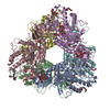
| ||||||||
|---|---|---|---|---|---|---|---|---|---|
| 1 |
| ||||||||
| Unit cell |
|
-Components
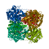
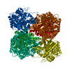
-Protein , 1 types, 6 molecules ABCDEF
| #1: Protein | Mass: 51249.449 Da / Num. of mol.: 6 Source method: isolated from a genetically manipulated source Source: (gene. exp.) Paenibacillus thiaminolyticus (bacteria)Gene: aLfuk1 / Plasmid: pET16b-alphaLF1 / Production host: |
|---|
-Sugars , 2 types, 14 molecules 
| #2: Polysaccharide | alpha-D-glucopyranose-(1-4)-alpha-D-glucopyranose / alpha-maltose   Source method: isolated from a genetically manipulated source Details: oligosaccharide / References: alpha-maltose #3: Sugar | ChemComp-GLC /  Type: D-saccharide, alpha linking / Mass: 180.156 Da / Num. of mol.: 6 Type: D-saccharide, alpha linking / Mass: 180.156 Da / Num. of mol.: 6Source method: isolated from a genetically manipulated source Formula: C6H12O6 |
|---|
-Non-polymers , 7 types, 1411 molecules 


| #4: Chemical | ChemComp-PGE /  Mass: 150.173 Da / Num. of mol.: 10 / Source method: obtained synthetically / Formula: C6H14O4 Mass: 150.173 Da / Num. of mol.: 10 / Source method: obtained synthetically / Formula: C6H14O4#5: Chemical |  Mass: 106.120 Da / Num. of mol.: 2 / Source method: obtained synthetically / Formula: C4H10O3 Mass: 106.120 Da / Num. of mol.: 2 / Source method: obtained synthetically / Formula: C4H10O3#6: Chemical |  Mass: 238.278 Da / Num. of mol.: 2 / Source method: obtained synthetically / Formula: C10H22O6 / Comment: precipitant*YM Mass: 238.278 Da / Num. of mol.: 2 / Source method: obtained synthetically / Formula: C10H22O6 / Comment: precipitant*YM#7: Chemical | ChemComp-PG4 / |  Mass: 194.226 Da / Num. of mol.: 1 / Source method: obtained synthetically / Formula: C8H18O5 / Comment: precipitant*YM Mass: 194.226 Da / Num. of mol.: 1 / Source method: obtained synthetically / Formula: C8H18O5 / Comment: precipitant*YM#8: Chemical |  Mass: 282.331 Da / Num. of mol.: 2 / Source method: obtained synthetically / Formula: C12H26O7 / Comment: precipitant*YM Mass: 282.331 Da / Num. of mol.: 2 / Source method: obtained synthetically / Formula: C12H26O7 / Comment: precipitant*YM#9: Chemical |  Mass: 22.990 Da / Num. of mol.: 2 / Source method: obtained synthetically / Formula: Na Mass: 22.990 Da / Num. of mol.: 2 / Source method: obtained synthetically / Formula: Na#10: Water | ChemComp-HOH / | Mass: 18.015 Da / Num. of mol.: 1392 / Source method: isolated from a natural source / Formula: H2O |
|---|
-Experimental details
-Experiment
| Experiment | Method: X-RAY DIFFRACTION / Number of used crystals: 1 |
|---|
- Sample preparation
| Crystal | Density Matthews: 2.54 Å3/Da / Density % sol: 51.7 % |
|---|---|
| Crystal grow | Temperature: 291.15 K / Method: vapor diffusion, sitting drop / pH: 6.5 Details: 25% (w/v) PEG 3350, 0.2 M Ammonium acetate, 0.1 M BIS-TRIS buffer, Additive: 50 mM Maltose |
-Data collection
| Diffraction | Mean temperature: 100 K |
|---|---|
| Diffraction source | Source: SYNCHROTRON / Site: PETRA III, EMBL c/o DESY  / Beamline: P13 (MX1) / Wavelength: 0.9201 Å / Beamline: P13 (MX1) / Wavelength: 0.9201 Å |
| Detector | Type: DECTRIS PILATUS 6M-F / Detector: PIXEL / Date: Sep 11, 2015 |
| Radiation | Protocol: SINGLE WAVELENGTH / Monochromatic (M) / Laue (L): M / Scattering type: x-ray |
| Radiation wavelength | Wavelength: 0.9201 Å / Relative weight: 1 |
| Reflection | Resolution: 2.2→48.93 Å / Num. obs: 157780 / % possible obs: 99.4 % / Observed criterion σ(I): -3.7 / Redundancy: 5 % / Biso Wilson estimate: 24.8 Å2 / Rmerge(I) obs: 0.099 / Net I/σ(I): 12.3 |
| Reflection shell | Resolution: 2.2→2.24 Å / Redundancy: 4.7 % / Rmerge(I) obs: 0.709 / Mean I/σ(I) obs: 2.1 / % possible all: 99.2 |
- Processing
| Software |
| ||||||||||||||||||||||||||||||||||||||||||||||||||||||||||||||||||||||||||||||||||||||||||||||||||||||||||||||||||||||||||||||||||||||||||||||||||||||||||||||||||||||||||||||||||||||
|---|---|---|---|---|---|---|---|---|---|---|---|---|---|---|---|---|---|---|---|---|---|---|---|---|---|---|---|---|---|---|---|---|---|---|---|---|---|---|---|---|---|---|---|---|---|---|---|---|---|---|---|---|---|---|---|---|---|---|---|---|---|---|---|---|---|---|---|---|---|---|---|---|---|---|---|---|---|---|---|---|---|---|---|---|---|---|---|---|---|---|---|---|---|---|---|---|---|---|---|---|---|---|---|---|---|---|---|---|---|---|---|---|---|---|---|---|---|---|---|---|---|---|---|---|---|---|---|---|---|---|---|---|---|---|---|---|---|---|---|---|---|---|---|---|---|---|---|---|---|---|---|---|---|---|---|---|---|---|---|---|---|---|---|---|---|---|---|---|---|---|---|---|---|---|---|---|---|---|---|---|---|---|---|
| Refinement | Method to determine structure: MOLECULAR REPLACEMENT Starting model: 2WVS Resolution: 2.2→48.93 Å / Cor.coef. Fo:Fc: 0.962 / SU B: 6.104 / SU ML: 0.134 / Cross valid method: FREE R-VALUE / ESU R: 0.214 Stereochemistry target values: THE STEREOCHEMISTRY LIBRARY OF CCP4 VERSION 7.0 Details: HYDROGENS HAVE BEEN ADDED IN THE RIDING POSITIONS. THE LAST REFINEMENT CYCLE WAS PERFORMED AGAINST ALL REFLECTIONS OF THE DATA SET.
| ||||||||||||||||||||||||||||||||||||||||||||||||||||||||||||||||||||||||||||||||||||||||||||||||||||||||||||||||||||||||||||||||||||||||||||||||||||||||||||||||||||||||||||||||||||||
| Solvent computation | Ion probe radii: 0.8 Å / Shrinkage radii: 0.8 Å / VDW probe radii: 1.2 Å | ||||||||||||||||||||||||||||||||||||||||||||||||||||||||||||||||||||||||||||||||||||||||||||||||||||||||||||||||||||||||||||||||||||||||||||||||||||||||||||||||||||||||||||||||||||||
| Displacement parameters | Biso mean: 33.97 Å2
| ||||||||||||||||||||||||||||||||||||||||||||||||||||||||||||||||||||||||||||||||||||||||||||||||||||||||||||||||||||||||||||||||||||||||||||||||||||||||||||||||||||||||||||||||||||||
| Refinement step | Cycle: LAST / Resolution: 2.2→48.93 Å
| ||||||||||||||||||||||||||||||||||||||||||||||||||||||||||||||||||||||||||||||||||||||||||||||||||||||||||||||||||||||||||||||||||||||||||||||||||||||||||||||||||||||||||||||||||||||
| Refine LS restraints |
|