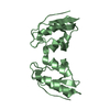
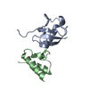
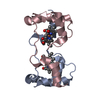
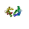
PROTEIN TRANSPORT / protein-protein complex / uim and ubiquitin
Function / homology
Function and homology information
Inhibition of membrane repair / ESCRT-0 complex / : / membrane invagination / Translesion synthesis by REV1 / Recognition of DNA damage by PCNA-containing replication complex / Translesion Synthesis by POLH / Downregulation of ERBB4 signaling / Spry regulation of FGF signaling / Downregulation of ERBB2:ERBB3 signaling ...Inhibition of membrane repair / ESCRT-0 complex / : / membrane invagination / Translesion synthesis by REV1 / Recognition of DNA damage by PCNA-containing replication complex / Translesion Synthesis by POLH / Downregulation of ERBB4 signaling / Spry regulation of FGF signaling / Downregulation of ERBB2:ERBB3 signaling / NOD1/2 Signaling Pathway / APC/C:Cdc20 mediated degradation of Cyclin B / SCF-beta-TrCP mediated degradation of Emi1 / APC-Cdc20 mediated degradation of Nek2A / EGFR downregulation / TCF dependent signaling in response to WNT / NRIF signals cell death from the nucleus / p75NTR recruits signalling complexes / NF-kB is activated and signals survival / Activated NOTCH1 Transmits Signal to the Nucleus / Downregulation of TGF-beta receptor signaling / TGF-beta receptor signaling in EMT (epithelial to mesenchymal transition) / Downregulation of SMAD2/3:SMAD4 transcriptional activity / SMAD2/SMAD3:SMAD4 heterotrimer regulates transcription / Senescence-Associated Secretory Phenotype (SASP) / Regulation of innate immune responses to cytosolic DNA / activated TAK1 mediates p38 MAPK activation / JNK (c-Jun kinases) phosphorylation and activation mediated by activated human TAK1 / Regulation of FZD by ubiquitination / PINK1-PRKN Mediated Mitophagy / N-glycan trimming in the ER and Calnexin/Calreticulin cycle / Regulation of TNFR1 signaling / TNFR1-induced NF-kappa-B signaling pathway / Translesion synthesis by POLK / Translesion synthesis by POLI / Regulation of necroptotic cell death / MAP3K8 (TPL2)-dependent MAPK1/3 activation / HDR through Homologous Recombination (HRR) / Josephin domain DUBs / Recruitment and ATM-mediated phosphorylation of repair and signaling proteins at DNA double strand breaks / DNA Damage Recognition in GG-NER / Formation of Incision Complex in GG-NER / Gap-filling DNA repair synthesis and ligation in GG-NER / Dual Incision in GG-NER / Fanconi Anemia Pathway / Regulation of TP53 Activity through Phosphorylation / Regulation of TP53 Degradation / Regulation of TP53 Activity through Methylation / Negative regulation of MET activity / Cyclin D associated events in G1 / PTK6 Regulates RTKs and Their Effectors AKT1 and DOK1 / Downregulation of ERBB2 signaling / E3 ubiquitin ligases ubiquitinate target proteins / Regulation of PTEN localization / ER Quality Control Compartment (ERQC) / Regulation of expression of SLITs and ROBOs / Interferon alpha/beta signaling / Endosomal Sorting Complex Required For Transport (ESCRT) / Activation of IRF3, IRF7 mediated by TBK1, IKKε (IKBKE) / IKK complex recruitment mediated by RIP1 / IRAK2 mediated activation of TAK1 complex / TRAF6-mediated induction of TAK1 complex within TLR4 complex / Alpha-protein kinase 1 signaling pathway / RAS processing / Pexophagy / Inactivation of CSF3 (G-CSF) signaling / Negative regulation of FLT3 / Regulation of BACH1 activity / IRAK2 mediated activation of TAK1 complex upon TLR7/8 or 9 stimulation / Regulation of NF-kappa B signaling / Termination of translesion DNA synthesis / Ovarian tumor domain proteases / Negative regulators of DDX58/IFIH1 signaling / Negative regulation of FGFR1 signaling / Negative regulation of FGFR2 signaling / Negative regulation of FGFR3 signaling / Negative regulation of FGFR4 signaling / Negative regulation of MAPK pathway / Synthesis of active ubiquitin: roles of E1 and E2 enzymes / Iron uptake and transport / Deactivation of the beta-catenin transactivating complex / Metalloprotease DUBs / Formation of TC-NER Pre-Incision Complex / Dual incision in TC-NER / Gap-filling DNA repair synthesis and ligation in TC-NER / Activation of NF-kappaB in B cells / L13a-mediated translational silencing of Ceruloplasmin expression / Autodegradation of Cdh1 by Cdh1:APC/C / APC/C:Cdc20 mediated degradation of Securin / APC/C:Cdh1 mediated degradation of Cdc20 and other APC/C:Cdh1 targeted proteins in late mitosis/early G1 / Cdc20:Phospho-APC/C mediated degradation of Cyclin A / SRP-dependent cotranslational protein targeting to membrane / SCF(Skp2)-mediated degradation of p27/p21 / FCERI mediated NF-kB activation / Autodegradation of the E3 ubiquitin ligase COP1 / Asymmetric localization of PCP proteins / Degradation of AXIN / Degradation of DVL / Hedgehog ligand biogenesis / Dectin-1 mediated noncanonical NF-kB signaling Similarity search - Function
Mass: 2388.556 Da / Num. of mol.: 1 / Source method: obtained synthetically Details: This sequence occurs naturally in human HRS and was synthesized by standard peptide synthesis methods. References: UniProt: O14964
Resolution: 1.7→36.51 Å / Cor.coef. Fo:Fc: 0.949 / Cor.coef. Fo:Fc free: 0.942 / SU B: 2.038 / SU ML: 0.069 / Cross valid method: THROUGHOUT / ESU R: 0.113 / ESU R Free: 0.109 / Stereochemistry target values: MAXIMUM LIKELIHOOD / Details: HYDROGENS HAVE BEEN ADDED IN THE RIDING POSITIONS
Rfactor
Num. reflection
% reflection
Selection details
Rfree
0.228
992
5.1 %
RANDOM
Rwork
0.197
-
-
-
all
0.1989
19706
-
-
obs
0.1989
19418
98.7 %
-
Solvent computation
Ion probe radii: 0.8 Å / Shrinkage radii: 0.8 Å / VDW probe radii: 1.4 Å / Solvent model: BABINET MODEL WITH MASK
Displacement parameters
Biso mean: 19.47 Å2
Baniso -1
Baniso -2
Baniso -3
1-
-0.58 Å2
-0.29 Å2
0 Å2
2-
-
-0.58 Å2
0 Å2
3-
-
-
0.88 Å2
Refinement step
Cycle: LAST / Resolution: 1.7→36.51 Å
Protein
Nucleic acid
Ligand
Solvent
Total
Num. atoms
1287
0
0
105
1392
Refine LS restraints
Refine-ID
Type
Dev ideal
Dev ideal target
Number
X-RAY DIFFRACTION
r_bond_refined_d
0.015
0.022
1301
X-RAY DIFFRACTION
r_bond_other_d
0.002
0.02
1219
X-RAY DIFFRACTION
r_angle_refined_deg
1.551
1.995
1750
X-RAY DIFFRACTION
r_angle_other_deg
0.788
3
2870
X-RAY DIFFRACTION
r_dihedral_angle_1_deg
5.594
5
159
X-RAY DIFFRACTION
r_dihedral_angle_2_deg
X-RAY DIFFRACTION
r_dihedral_angle_3_deg
X-RAY DIFFRACTION
r_dihedral_angle_4_deg
X-RAY DIFFRACTION
r_chiral_restr
0.09
0.2
211
X-RAY DIFFRACTION
r_gen_planes_refined
0.006
0.02
1401
X-RAY DIFFRACTION
r_gen_planes_other
0.002
0.02
209
X-RAY DIFFRACTION
r_nbd_refined
0.208
0.2
214
X-RAY DIFFRACTION
r_nbd_other
0.245
0.2
1419
X-RAY DIFFRACTION
r_nbtor_refined
X-RAY DIFFRACTION
r_nbtor_other
0.079
0.2
814
X-RAY DIFFRACTION
r_xyhbond_nbd_refined
0.143
0.2
90
X-RAY DIFFRACTION
r_xyhbond_nbd_other
X-RAY DIFFRACTION
r_metal_ion_refined
X-RAY DIFFRACTION
r_metal_ion_other
X-RAY DIFFRACTION
r_symmetry_vdw_refined
0.135
0.2
13
X-RAY DIFFRACTION
r_symmetry_vdw_other
0.303
0.2
59
X-RAY DIFFRACTION
r_symmetry_hbond_refined
0.133
0.2
18
X-RAY DIFFRACTION
r_symmetry_hbond_other
X-RAY DIFFRACTION
r_symmetry_metal_ion_refined
X-RAY DIFFRACTION
r_symmetry_metal_ion_other
X-RAY DIFFRACTION
r_mcbond_it
1.137
1.5
808
X-RAY DIFFRACTION
r_mcbond_other
X-RAY DIFFRACTION
r_mcangle_it
2.106
2
1313
X-RAY DIFFRACTION
r_scbond_it
3.145
3
493
X-RAY DIFFRACTION
r_scangle_it
5.455
4.5
437
X-RAY DIFFRACTION
r_rigid_bond_restr
X-RAY DIFFRACTION
r_sphericity_free
X-RAY DIFFRACTION
r_sphericity_bonded
LS refinement shell
Resolution: 1.7→1.74 Å / Total num. of bins used: 20 /
Rfactor
Num. reflection
Rfree
0.27
80
Rwork
0.228
1277
+
About Yorodumi
-
News
-
Feb 9, 2022. New format data for meta-information of EMDB entries
New format data for meta-information of EMDB entries
Version 3 of the EMDB header file is now the official format.
The previous official version 1.9 will be removed from the archive.
In the structure databanks used in Yorodumi, some data are registered as the other names, "COVID-19 virus" and "2019-nCoV". Here are the details of the virus and the list of structure data.
Jan 31, 2019. EMDB accession codes are about to change! (news from PDBe EMDB page)
EMDB accession codes are about to change! (news from PDBe EMDB page)
The allocation of 4 digits for EMDB accession codes will soon come to an end. Whilst these codes will remain in use, new EMDB accession codes will include an additional digit and will expand incrementally as the available range of codes is exhausted. The current 4-digit format prefixed with “EMD-” (i.e. EMD-XXXX) will advance to a 5-digit format (i.e. EMD-XXXXX), and so on. It is currently estimated that the 4-digit codes will be depleted around Spring 2019, at which point the 5-digit format will come into force.
The EM Navigator/Yorodumi systems omit the EMD- prefix.
Related info.:Q: What is EMD? / ID/Accession-code notation in Yorodumi/EM Navigator
Yorodumi is a browser for structure data from EMDB, PDB, SASBDB, etc.
This page is also the successor to EM Navigator detail page, and also detail information page/front-end page for Omokage search.
The word "yorodu" (or yorozu) is an old Japanese word meaning "ten thousand". "mi" (miru) is to see.
Related info.:EMDB / PDB / SASBDB / Comparison of 3 databanks / Yorodumi Search / Aug 31, 2016. New EM Navigator & Yorodumi / Yorodumi Papers / Jmol/JSmol / Function and homology information / Changes in new EM Navigator and Yorodumi
 Movie
Movie Controller


 Open data
 Basic information
 Components
 Keywords
 PROTEIN TRANSPORT / protein-protein complex / uim and ubiquitin
 Function and homology information
 Authors
 Citation
 Structure visualization
 Downloads & links
 Other downloads

 PDBj
PDBj


 Assembly
 Mass: 18.015 Da / Num. of mol.: 105 / Source method: isolated from a natural source / Formula: H2O
 Sample preparation
 / Beamline: AR-NW12A / Wavelength: 1 / Wavelength: 1 Å
 Processing