 Movie
Movie Controller


+ Open data

- Basic information
| Entry | Database: PDB / ID: 2cxn | ||||||
|---|---|---|---|---|---|---|---|
| Title | Crystal structure of mouse AMF / phosphate complex | ||||||
 Components Components | Glucose-6-phosphate isomerase | ||||||
 Keywords Keywords | ISOMERASE | ||||||
| Function / homology |  Function and homology information Function and homology informationglycolytic process through glucose-6-phosphate / Gluconeogenesis / Glycolysis / TP53 Regulates Metabolic Genes / glucose-6-phosphate isomerase / glucose-6-phosphate isomerase activity / glucose 6-phosphate metabolic process / carbohydrate derivative binding / fructose 6-phosphate metabolic process / monosaccharide binding ...glycolytic process through glucose-6-phosphate / Gluconeogenesis / Glycolysis / TP53 Regulates Metabolic Genes / glucose-6-phosphate isomerase / glucose-6-phosphate isomerase activity / glucose 6-phosphate metabolic process / carbohydrate derivative binding / fructose 6-phosphate metabolic process / monosaccharide binding / canonical glycolysis / positive regulation of immunoglobulin production / ciliary membrane / erythrocyte homeostasis / response to testosterone / response to immobilization stress / mesoderm formation / response to cadmium ion / response to muscle stretch / Neutrophil degranulation / positive regulation of endothelial cell migration / response to progesterone / cytokine activity / glycolytic process / gluconeogenesis / growth factor activity / response to estradiol / myelin sheath / glucose homeostasis / in utero embryonic development / learning or memory / ubiquitin protein ligase binding / negative regulation of apoptotic process / extracellular space / plasma membrane / cytosol Similarity search - Function | ||||||
| Biological species |  | ||||||
| Method |  X-RAY DIFFRACTION / SYNCHROTRON / FOURIER SYNTHESIS / Resolution: 1.4 Å X-RAY DIFFRACTION / SYNCHROTRON / FOURIER SYNTHESIS / Resolution: 1.4 Å | ||||||
 Authors Authors | Tanaka, N. / Haga, A. / Naba, N. / Shiraiwa, K. / Kusakabe, Y. / Hashimoto, K. / Funasaka, T. / Nagase, H. / Raz, A. / Nakamura, K.T. | ||||||
 Citation Citation | Journal: J.Mol.Biol. / Year: 2006 Title: Crystal structures of mouse autocrine motility factor in complex with carbohydrate phosphate inhibitors provide insight into structure-activity relationship of the inhibitors Authors: Tanaka, N. / Haga, A. / Naba, N. / Shiraiwa, K. / Kusakabe, Y. / Hashimoto, K. / Funasaka, T. / Nagase, H. / Raz, A. / Nakamura, K.T. | ||||||
| History |
|
- Structure visualization
| Structure viewer | Molecule: MolmilJmol/JSmol |
|---|
- Downloads & links
-Download
| PDBx/mmCIF format | 2cxn.cif.gz | 251.3 KB | Display | PDBx/mmCIF format |
|---|---|---|---|---|
| PDB format | pdb2cxn.ent.gz | 199.1 KB | Display | PDB format |
| PDBx/mmJSON format | 2cxn.json.gz | Tree view | PDBx/mmJSON format | |
| Others |  Other downloads Other downloads |
-Validation report
| Arichive directory | https://data.pdbj.org/pub/pdb/validation_reports/cx/2cxnftp://data.pdbj.org/pub/pdb/validation_reports/cx/2cxn | HTTPS FTP |
|---|
-Related structure data
| Related structure data |  2cvpC  2cxoC  2cxpC  2cxqC  2cxrC  2cxsC  2cxtC  2cxuC C: citing same article ( |
|---|---|
| Similar structure data |


-Links
 PDBj
PDBj


- Assembly
| Deposited unit | 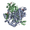
| ||||||||
|---|---|---|---|---|---|---|---|---|---|
| 1 |
| ||||||||
| Unit cell |
|
-Components
| #1: Protein | Mass: 62693.582 Da / Num. of mol.: 2 Source method: isolated from a genetically manipulated source Source: (gene. exp.)  #2: Chemical | ChemComp-PO4 / |   Mass: 94.971 Da / Num. of mol.: 1 / Source method: obtained synthetically / Formula: PO4 Mass: 94.971 Da / Num. of mol.: 1 / Source method: obtained synthetically / Formula: PO4#3: Chemical | ChemComp-GOL /   Mass: 92.094 Da / Num. of mol.: 4 / Source method: obtained synthetically / Formula: C3H8O3 Mass: 92.094 Da / Num. of mol.: 4 / Source method: obtained synthetically / Formula: C3H8O3#4: Water | ChemComp-HOH / |  Mass: 18.015 Da / Num. of mol.: 1192 / Source method: isolated from a natural source / Formula: H2O Mass: 18.015 Da / Num. of mol.: 1192 / Source method: isolated from a natural source / Formula: H2O |
|---|
-Experimental details
-Experiment
| Experiment | Method: X-RAY DIFFRACTION / Number of used crystals: 1 |
|---|
- Sample preparation
| Crystal | Density Matthews: 2.32 Å3/Da / Density % sol: 47 % |
|---|---|
| Crystal grow | Temperature: 293 K / Method: vapor diffusion, hanging drop / pH: 7.5 Details: Cacodylate, PEG8000, NaAcetate, pH 7.5, VAPOR DIFFUSION, HANGING DROP, temperature 293K |
-Data collection
| Diffraction | Mean temperature: 100 K |
|---|---|
| Diffraction source | Source: SYNCHROTRON / Site: Photon Factory  / Beamline: BL-5A / Wavelength: 1 Å / Beamline: BL-5A / Wavelength: 1 Å |
| Detector | Type: ADSC QUANTUM 315 / Detector: CCD |
| Radiation | Protocol: SINGLE WAVELENGTH / Monochromatic (M) / Laue (L): M / Scattering type: x-ray |
| Radiation wavelength | Wavelength: 1 Å / Relative weight: 1 |
| Reflection | Resolution: 1.4→40 Å / Num. obs: 215161 / % possible obs: 96.1 % |
- Processing
| Software |
| ||||||||||||||||||||||||||||||||||||||||||||||||||||||||||||||||||||||||||||||||||||||||||||||||||||||||||||||||||||||||||||||||||||||||||||||||||||||||||||||||||||||||||
|---|---|---|---|---|---|---|---|---|---|---|---|---|---|---|---|---|---|---|---|---|---|---|---|---|---|---|---|---|---|---|---|---|---|---|---|---|---|---|---|---|---|---|---|---|---|---|---|---|---|---|---|---|---|---|---|---|---|---|---|---|---|---|---|---|---|---|---|---|---|---|---|---|---|---|---|---|---|---|---|---|---|---|---|---|---|---|---|---|---|---|---|---|---|---|---|---|---|---|---|---|---|---|---|---|---|---|---|---|---|---|---|---|---|---|---|---|---|---|---|---|---|---|---|---|---|---|---|---|---|---|---|---|---|---|---|---|---|---|---|---|---|---|---|---|---|---|---|---|---|---|---|---|---|---|---|---|---|---|---|---|---|---|---|---|---|---|---|---|---|---|---|
| Refinement | Method to determine structure: FOURIER SYNTHESIS / Resolution: 1.4→40 Å / Cor.coef. Fo:Fc: 0.964 / Cor.coef. Fo:Fc free: 0.957 / SU B: 0.902 / SU ML: 0.036 / Cross valid method: THROUGHOUT / ESU R: 0.061 / ESU R Free: 0.06 / Stereochemistry target values: MAXIMUM LIKELIHOOD
| ||||||||||||||||||||||||||||||||||||||||||||||||||||||||||||||||||||||||||||||||||||||||||||||||||||||||||||||||||||||||||||||||||||||||||||||||||||||||||||||||||||||||||
| Solvent computation | Ion probe radii: 0.8 Å / Shrinkage radii: 0.8 Å / VDW probe radii: 1.4 Å / Solvent model: BABINET MODEL WITH MASK | ||||||||||||||||||||||||||||||||||||||||||||||||||||||||||||||||||||||||||||||||||||||||||||||||||||||||||||||||||||||||||||||||||||||||||||||||||||||||||||||||||||||||||
| Displacement parameters | Biso mean: 14.502 Å2
| ||||||||||||||||||||||||||||||||||||||||||||||||||||||||||||||||||||||||||||||||||||||||||||||||||||||||||||||||||||||||||||||||||||||||||||||||||||||||||||||||||||||||||
| Refinement step | Cycle: LAST / Resolution: 1.4→40 Å
| ||||||||||||||||||||||||||||||||||||||||||||||||||||||||||||||||||||||||||||||||||||||||||||||||||||||||||||||||||||||||||||||||||||||||||||||||||||||||||||||||||||||||||
| Refine LS restraints |
| ||||||||||||||||||||||||||||||||||||||||||||||||||||||||||||||||||||||||||||||||||||||||||||||||||||||||||||||||||||||||||||||||||||||||||||||||||||||||||||||||||||||||||
| LS refinement shell | Resolution: 1.4→1.436 Å / Total num. of bins used: 20 /
|
