 Movie
Movie Controller


+ Open data

- Basic information
| Entry | Database: PDB / ID: 2bsq | ||||||
|---|---|---|---|---|---|---|---|
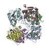
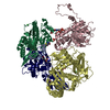
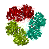
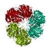
| Title | FitAB bound to DNA | ||||||
 Components Components |
| ||||||
 Keywords Keywords | TRANSCRIPTION / TRANSCRIPTION REGULATION COMPLEX / PIN DOMAIN / RIBBON-HELIX-HELIX / DNA BINDING / HETERODIMER | ||||||
| Function / homology |  Function and homology information Function and homology informationmigration in host / RNA nuclease activity / sequence-specific DNA binding / Hydrolases; Acting on ester bonds / regulation of DNA-templated transcription / magnesium ion binding / protein homodimerization activity / DNA binding Similarity search - Function | ||||||
| Biological species |  NEISSERIA GONORRHOEAE (bacteria) NEISSERIA GONORRHOEAE (bacteria) | ||||||
| Method |  X-RAY DIFFRACTION / SYNCHROTRON / MOLECULAR REPLACEMENT / Resolution: 3 Å X-RAY DIFFRACTION / SYNCHROTRON / MOLECULAR REPLACEMENT / Resolution: 3 Å | ||||||
 Authors Authors | Mattison, K. / Wilbur, J.S. / So, M. / Brennan, R.G. | ||||||
 Citation Citation | Journal: J.Biol.Chem. / Year: 2006 Title: Structure of Fitab from Neisseria Gonorrhoeae Bound to DNA Reveals a Tetramer of Toxin-Antitoxin Heterodimers Containing Pin Domains and Ribbon-Helix-Helix Motifs. Authors: Mattison, K. / Wilbur, J.S. / So, M. / Brennan, R.G. | ||||||
| History |
|
- Structure visualization
| Structure viewer | Molecule: MolmilJmol/JSmol |
|---|
- Downloads & links
-Download
| PDBx/mmCIF format | 2bsq.cif.gz | 208.9 KB | Display | PDBx/mmCIF format |
|---|---|---|---|---|
| PDB format | pdb2bsq.ent.gz | 162.3 KB | Display | PDB format |
| PDBx/mmJSON format | 2bsq.json.gz | Tree view | PDBx/mmJSON format | |
| Others |  Other downloads Other downloads |
-Validation report
| Summary document | 2bsq_validation.pdf.gz | 500 KB | Display | wwPDB validaton report |
|---|---|---|---|---|
| Full document | 2bsq_full_validation.pdf.gz | 538.2 KB | Display | |
| Data in XML | 2bsq_validation.xml.gz | 36.5 KB | Display | |
| Data in CIF | 2bsq_validation.cif.gz | 50.4 KB | Display | |
| Arichive directory | https://data.pdbj.org/pub/pdb/validation_reports/bs/2bsqftp://data.pdbj.org/pub/pdb/validation_reports/bs/2bsq | HTTPS FTP |
-Related structure data
| Related structure data |  2h1cC 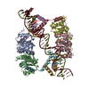 2h1oC  1yh4 S: Starting model for refinement C: citing same article ( |
|---|---|
| Similar structure data |


-Links
 PDBj
PDBj


- Assembly
| Deposited unit | 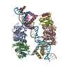
| ||||||||
|---|---|---|---|---|---|---|---|---|---|
| 1 |
| ||||||||
| Unit cell |
|
-Components
| #1: Protein | Mass: 16333.761 Da / Num. of mol.: 4 / Fragment: PIN DOMAIN, RESIDUES 1-139 / Mutation: YES Source method: isolated from a genetically manipulated source Source: (gene. exp.) NEISSERIA GONORRHOEAE (bacteria) / Plasmid: PET28B / Production host: #2: Protein | Mass: 8313.364 Da / Num. of mol.: 4 / Fragment: DNA-BINDING PROTEIN, RESIDUES 2-78 Source method: isolated from a genetically manipulated source Source: (gene. exp.) NEISSERIA GONORRHOEAE (bacteria) / Plasmid: PET28B / Production host: #3: DNA chain | | Mass: 11169.996 Da / Num. of mol.: 1 / Source method: obtained synthetically / Source: (synth.) NEISSERIA GONORRHOEAE (bacteria)#4: DNA chain | | Mass: 11198.117 Da / Num. of mol.: 1 / Source method: obtained synthetically / Source: (synth.) NEISSERIA GONORRHOEAE (bacteria)#5: Water | ChemComp-HOH / |  Mass: 18.015 Da / Num. of mol.: 45 / Source method: isolated from a natural source / Formula: H2O Mass: 18.015 Da / Num. of mol.: 45 / Source method: isolated from a natural source / Formula: H2OCompound details | ENGINEERED RESIDUE IN CHAIN A, ASP 139 TO LEU ENGINEERED RESIDUE IN CHAIN B, ASP 139 TO LEU ...ENGINEERED | |
|---|
-Experimental details
-Experiment
| Experiment | Method: X-RAY DIFFRACTION / Number of used crystals: 1 |
|---|
- Sample preparation
| Crystal | Density Matthews: 3.45 Å3/Da / Density % sol: 67.38 % Description: MODEL FILE NOT YET RELEASED-WILL BE IN SAME PAPER |
|---|---|
| Crystal grow | pH: 4.5 Details: 0.1 M ACETATE, PH 4.0 7.2 % PEG 20,000 7.2 % PEG 550 MME 0.26 M NA ACETATE, PH 7.0 |
-Data collection
| Diffraction | Mean temperature: 100 K |
|---|---|
| Diffraction source | Source: SYNCHROTRON / Site: ALS  / Beamline: 8.2.1 / Wavelength: 1.0332 / Beamline: 8.2.1 / Wavelength: 1.0332 |
| Detector | Type: ADSC CCD / Detector: CCD / Date: Mar 8, 2005 |
| Radiation | Monochromator: DOUBLE CRYSTAL, SI(111) / Protocol: SINGLE WAVELENGTH / Monochromatic (M) / Laue (L): M / Scattering type: x-ray |
| Radiation wavelength | Wavelength: 1.0332 Å / Relative weight: 1 |
| Reflection | Resolution: 3→81.65 Å / Num. obs: 16708 / % possible obs: 99.9 % / Observed criterion σ(I): 1.5 / Redundancy: 5.4 % / Rmerge(I) obs: 0.18 / Net I/σ(I): 3.1 |
| Reflection shell | Resolution: 3→3.15 Å / Redundancy: 5.4 % / Rmerge(I) obs: 0.44 / Mean I/σ(I) obs: 1.5 / % possible all: 99.9 |
- Processing
| Software |
| ||||||||||||||||||||||||||||||||||||||||||||||||||||||||||||||||||||||||||||||||
|---|---|---|---|---|---|---|---|---|---|---|---|---|---|---|---|---|---|---|---|---|---|---|---|---|---|---|---|---|---|---|---|---|---|---|---|---|---|---|---|---|---|---|---|---|---|---|---|---|---|---|---|---|---|---|---|---|---|---|---|---|---|---|---|---|---|---|---|---|---|---|---|---|---|---|---|---|---|---|---|---|---|
| Refinement | Method to determine structure: MOLECULAR REPLACEMENT Starting model: PDB ENTRY 1YH4 1yh4 Resolution: 3→70.36 Å / Rfactor Rfree error: 0.005 / Data cutoff high absF: 1657362.62 / Isotropic thermal model: RESTRAINED / Cross valid method: THROUGHOUT / σ(F): 1.5 / Stereochemistry target values: MAXIMUM LIKELIHOOD
| ||||||||||||||||||||||||||||||||||||||||||||||||||||||||||||||||||||||||||||||||
| Solvent computation | Solvent model: FLAT MODEL / Bsol: 26.4919 Å2 / ksol: 0.315881 e/Å3 | ||||||||||||||||||||||||||||||||||||||||||||||||||||||||||||||||||||||||||||||||
| Displacement parameters | Biso mean: 40.7 Å2
| ||||||||||||||||||||||||||||||||||||||||||||||||||||||||||||||||||||||||||||||||
| Refine analyze |
| ||||||||||||||||||||||||||||||||||||||||||||||||||||||||||||||||||||||||||||||||
| Refinement step | Cycle: LAST / Resolution: 3→70.36 Å
| ||||||||||||||||||||||||||||||||||||||||||||||||||||||||||||||||||||||||||||||||
| Refine LS restraints |
| ||||||||||||||||||||||||||||||||||||||||||||||||||||||||||||||||||||||||||||||||
| LS refinement shell | Resolution: 3→3.19 Å / Rfactor Rfree error: 0.017 / Total num. of bins used: 6
| ||||||||||||||||||||||||||||||||||||||||||||||||||||||||||||||||||||||||||||||||
| Xplor file |
|
