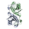
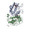
migration in host / RNA nuclease activity / sequence-specific DNA binding / Hydrolases; Acting on ester bonds / hydrolase activity / regulation of DNA-templated transcription / magnesium ion binding / protein homodimerization activity / DNA binding Similarity search - Function
Mass: 18.015 Da / Num. of mol.: 89 / Source method: isolated from a natural source / Formula: H2O
-
Experimental details
-
Experiment
Experiment
Method: X-RAY DIFFRACTION / Number of used crystals: 1
-
Sample preparation
Crystal
Density Matthews: 2.18 Å3/Da / Density % sol: 43.56 %
Crystal grow
Temperature: 298 K / Method: vapor diffusion, hanging drop / pH: 4.7 Details: 0.26 M sodium phosphate/citrate pH 4.7 and 2.0 M ammonium sulphate, VAPOR DIFFUSION, HANGING DROP, temperature 298K
-
Data collection
Diffraction
ID
Mean temperature (K)
Crystal-ID
1
100
1
2
100
1
1,2
1
Diffraction source
Source
Site
Beamline
ID
Wavelength (Å)
SYNCHROTRON
ALS
8.2.1
1
1
SYNCHROTRON
ALS
8.2.1
2
0.9796, 0.9686, 0.9794
Detector
Type
ID
Detector
Date
ADSC QUANTUM 210
1
CCD
Mar 8, 2005
ADSC QUANTUM 210
2
CCD
Mar 8, 2005
Radiation
ID
Monochromator
Protocol
Monochromatic (M) / Laue (L)
Scattering type
Wavelength-ID
1
Doublecrystal, Si(111)
SINGLEWAVELENGTH
M
x-ray
1
2
Doublecrystal, Si(111)
MAD
M
x-ray
1
Radiation wavelength
ID
Wavelength (Å)
Relative weight
1
1
1
2
0.9796
1
3
0.9686
1
4
0.9794
1
Reflection
Resolution: 1.8→39.11 Å / Num. all: 31002 / Num. obs: 13340 / % possible obs: 96.5 % / Observed criterion σ(F): 0 / Observed criterion σ(I): 9.7 / Biso Wilson estimate: 18.8 Å2 / Limit h max: 34 / Limit h min: -38 / Limit k max: 28 / Limit k min: -38 / Limit l max: 26 / Limit l min: 0 / Observed criterion F max: 855178.06 / Observed criterion F min: 6.8 / Rsym value: 0.055
Reflection shell
Resolution: 1.8→1.9 Å / Mean I/σ(I) obs: 3.1 / Num. unique all: 13340 / Rsym value: 0.222 / % possible all: 95.3
In the structure databanks used in Yorodumi, some data are registered as the other names, "COVID-19 virus" and "2019-nCoV". Here are the details of the virus and the list of structure data.
Jan 31, 2019. EMDB accession codes are about to change! (news from PDBe EMDB page)
EMDB accession codes are about to change! (news from PDBe EMDB page)
The allocation of 4 digits for EMDB accession codes will soon come to an end. Whilst these codes will remain in use, new EMDB accession codes will include an additional digit and will expand incrementally as the available range of codes is exhausted. The current 4-digit format prefixed with “EMD-” (i.e. EMD-XXXX) will advance to a 5-digit format (i.e. EMD-XXXXX), and so on. It is currently estimated that the 4-digit codes will be depleted around Spring 2019, at which point the 5-digit format will come into force.
The EM Navigator/Yorodumi systems omit the EMD- prefix.
Related info.:Q: What is EMD? / ID/Accession-code notation in Yorodumi/EM Navigator
Yorodumi is a browser for structure data from EMDB, PDB, SASBDB, etc.
This page is also the successor to EM Navigator detail page, and also detail information page/front-end page for Omokage search.
The word "yorodu" (or yorozu) is an old Japanese word meaning "ten thousand". "mi" (miru) is to see.
Related info.:EMDB / PDB / SASBDB / Comparison of 3 databanks / Yorodumi Search / Aug 31, 2016. New EM Navigator & Yorodumi / Yorodumi Papers / Jmol/JSmol / Function and homology information / Changes in new EM Navigator and Yorodumi
 Movie
Movie Controller


 Open data
 Basic information
 Components
 Keywords
 Function and homology information
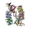
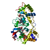
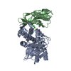
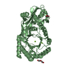
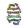
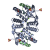
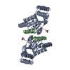
 Neisseria gonorrhoeae (bacteria)
 X-RAY DIFFRACTION /
  Authors
 Citation
 Structure visualization
 Downloads & links
 Other downloads
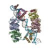


 PDBj
PDBj

 Assembly
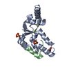


 Mass: 59.044 Da / Num. of mol.: 1 / Source method: obtained synthetically / Formula: C2H3O2
 Mass: 96.063 Da / Num. of mol.: 2 / Source method: obtained synthetically / Formula: SO4
 Mass: 24.305 Da / Num. of mol.: 3 / Source method: obtained synthetically / Formula: Mg
 Sample preparation
 Processing