 Movie
Movie Controller


+ Open data

- Basic information
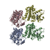
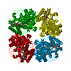
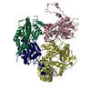
| Entry | Database: PDB / ID: 2h1o | ||||||
|---|---|---|---|---|---|---|---|
| Title | Structure of FitAB bound to IR36 DNA fragment | ||||||
 Components Components |
| ||||||
 Keywords Keywords | GENE REGULATION/DNA COMPLEX / PIN domain / RHH protein / DNA binding / tetramer of dimers / GENE REGULATION-DNA COMPLEX COMPLEX | ||||||
| Function / homology |  Function and homology information Function and homology informationmigration in host / RNA nuclease activity / sequence-specific DNA binding / Hydrolases; Acting on ester bonds / hydrolase activity / regulation of DNA-templated transcription / magnesium ion binding / protein homodimerization activity / DNA binding Similarity search - Function | ||||||
| Biological species |  Neisseria gonorrhoeae (bacteria) Neisseria gonorrhoeae (bacteria) | ||||||
| Method |  X-RAY DIFFRACTION / SYNCHROTRON / MOLECULAR REPLACEMENT / Resolution: 3 Å X-RAY DIFFRACTION / SYNCHROTRON / MOLECULAR REPLACEMENT / Resolution: 3 Å | ||||||
 Authors Authors | Mattison, K. / Wilbur, J.S. / So, M. / Brennan, R.G. | ||||||
 Citation Citation | Journal: J.Biol.Chem. / Year: 2006 Title: Structure of FitAB from Neisseria gonorrhoeae bound to DNA reveals a tetramer of toxin-antitoxin heterodimers containing pin domains and ribbon-helix-helix motifs. Authors: Mattison, K. / Wilbur, J.S. / So, M. / Brennan, R.G. | ||||||
| History |
|
- Structure visualization
| Structure viewer | Molecule: MolmilJmol/JSmol |
|---|
- Downloads & links
-Download
| PDBx/mmCIF format | 2h1o.cif.gz | 208 KB | Display | PDBx/mmCIF format |
|---|---|---|---|---|
| PDB format | pdb2h1o.ent.gz | 161.7 KB | Display | PDB format |
| PDBx/mmJSON format | 2h1o.json.gz | Tree view | PDBx/mmJSON format | |
| Others |  Other downloads Other downloads |
-Validation report
| Arichive directory | https://data.pdbj.org/pub/pdb/validation_reports/h1/2h1oftp://data.pdbj.org/pub/pdb/validation_reports/h1/2h1o | HTTPS FTP |
|---|
-Related structure data
| Related structure data | 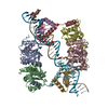 2bsqC  2h1cSC S: Starting model for refinement C: citing same article ( |
|---|---|
| Similar structure data |


-Links
 PDBj
PDBj


- Assembly
| Deposited unit | 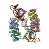
| ||||||||
|---|---|---|---|---|---|---|---|---|---|
| 1 |
| ||||||||
| Unit cell |
|
-Components
| #1: DNA chain | Mass: 11169.996 Da / Num. of mol.: 1 / Mutation: iodo / Source method: obtained synthetically / Details: sequence upstream of FitAB promoter region | ||||
|---|---|---|---|---|---|
| #2: DNA chain | Mass: 11198.117 Da / Num. of mol.: 1 / Mutation: iodo / Source method: obtained synthetically / Details: sequence upstream of FitAB promoter region | ||||
| #3: Protein | Mass: 15919.320 Da / Num. of mol.: 4 Source method: isolated from a genetically manipulated source Source: (gene. exp.) Neisseria gonorrhoeae (bacteria) / Gene: fitB / Species (production host): Escherichia coli / Production host: #4: Protein | Mass: 7283.293 Da / Num. of mol.: 4 Source method: isolated from a genetically manipulated source Source: (gene. exp.) Neisseria gonorrhoeae (bacteria) / Gene: fitA / Species (production host): Escherichia coli / Production host: #5: Water | ChemComp-HOH / |  Mass: 18.015 Da / Num. of mol.: 46 / Source method: isolated from a natural source / Formula: H2O Mass: 18.015 Da / Num. of mol.: 46 / Source method: isolated from a natural source / Formula: H2O |
-Experimental details
-Experiment
| Experiment | Method: X-RAY DIFFRACTION / Number of used crystals: 1 |
|---|
- Sample preparation
| Crystal | Density Matthews: 3.61 Å3/Da / Density % sol: 65.94 % | ||||||||||||||||||||||||||||||||
|---|---|---|---|---|---|---|---|---|---|---|---|---|---|---|---|---|---|---|---|---|---|---|---|---|---|---|---|---|---|---|---|---|---|
| Crystal grow | Temperature: 298 K / Method: vapor diffusion, hanging drop / pH: 5.6 Details: 0.1 M sodium acetate, pH 4.0, 0.27 M sodium acetate, pH 7.0, 7.2 % PEG 20,000, 7.2 % PEG monomethyl ether 550, pH 5.6, VAPOR DIFFUSION, HANGING DROP, temperature 298K | ||||||||||||||||||||||||||||||||
| Components of the solutions |
|
-Data collection
| Diffraction | Mean temperature: 100 K |
|---|---|
| Diffraction source | Source: SYNCHROTRON / Site: ALS  / Beamline: 8.2.1 / Wavelength: 1.0332 Å / Beamline: 8.2.1 / Wavelength: 1.0332 Å |
| Detector | Type: ADSC QUANTUM 210 / Detector: CCD / Date: Mar 8, 2005 |
| Radiation | Monochromator: Double crystal, Si(111) / Protocol: SINGLE WAVELENGTH / Monochromatic (M) / Laue (L): M / Scattering type: x-ray |
| Radiation wavelength | Wavelength: 1.0332 Å / Relative weight: 1 |
| Reflection | Resolution: 2.99→70.36 Å / Num. all: 33360 / Num. obs: 33243 / % possible obs: 99.9 % / Observed criterion σ(F): 0 / Observed criterion σ(I): 1.5 / Rsym value: 0.178 / Net I/σ(I): 3.1 |
| Reflection shell | Resolution: 2.99→3.14 Å / Mean I/σ(I) obs: 44 / Rsym value: 0.015 / % possible all: 99.9 |
- Processing
| Software |
| ||||||||||||||||||||||||||||||||||||||||||||||||||||||||||||||||||||||||||||||||||||||||||
|---|---|---|---|---|---|---|---|---|---|---|---|---|---|---|---|---|---|---|---|---|---|---|---|---|---|---|---|---|---|---|---|---|---|---|---|---|---|---|---|---|---|---|---|---|---|---|---|---|---|---|---|---|---|---|---|---|---|---|---|---|---|---|---|---|---|---|---|---|---|---|---|---|---|---|---|---|---|---|---|---|---|---|---|---|---|---|---|---|---|---|---|
| Refinement | Method to determine structure: MOLECULAR REPLACEMENT Starting model: PDB entry 2H1C Resolution: 3→70.36 Å / Rfactor Rfree error: 0.005 / Occupancy max: 1 / Occupancy min: 1 / Cross valid method: THROUGHOUT / σ(F): 0 / Stereochemistry target values: Engh & Huber
| ||||||||||||||||||||||||||||||||||||||||||||||||||||||||||||||||||||||||||||||||||||||||||
| Solvent computation | Solvent model: CNS bulk solvent model used / Bsol: 25.6821 Å2 / ksol: 0.316642 e/Å3 | ||||||||||||||||||||||||||||||||||||||||||||||||||||||||||||||||||||||||||||||||||||||||||
| Displacement parameters | Biso mean: 40.87 Å2
| ||||||||||||||||||||||||||||||||||||||||||||||||||||||||||||||||||||||||||||||||||||||||||
| Refine analyze |
| ||||||||||||||||||||||||||||||||||||||||||||||||||||||||||||||||||||||||||||||||||||||||||
| Refinement step | Cycle: LAST / Resolution: 3→70.36 Å
| ||||||||||||||||||||||||||||||||||||||||||||||||||||||||||||||||||||||||||||||||||||||||||
| Refine LS restraints |
| ||||||||||||||||||||||||||||||||||||||||||||||||||||||||||||||||||||||||||||||||||||||||||
| LS refinement shell | Refine-ID: X-RAY DIFFRACTION
|
