 Movie
Movie Controller

[English] 日本語
 Yorodumi
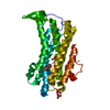
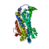
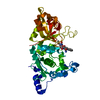
- PDB-1z8g: Crystal structure of the extracellular region of the transmembran... -
+ Open data

- Basic information
| Entry | Database: PDB / ID: 1z8g | ||||||
|---|---|---|---|---|---|---|---|
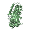
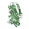
| Title | Crystal structure of the extracellular region of the transmembrane serine protease hepsin with covalently bound preferred substrate. | ||||||
 Components Components |
| ||||||
 Keywords Keywords | HYDROLASE/HYDROLASE INHIBITOR / SERINE PROTEASE HEPSIN / PROTEASE / HYDROLASE-HYDROLASE INHIBITOR COMPLEX | ||||||
| Function / homology |  Function and homology information Function and homology informationhepsin / pilomotor reflex / positive regulation of thyroid hormone generation / Signaling by MST1 / serine-type exopeptidase activity / positive regulation of plasminogen activation / basement membrane disassembly / response to thyroid hormone / cochlea morphogenesis / MET Receptor Activation ...hepsin / pilomotor reflex / positive regulation of thyroid hormone generation / Signaling by MST1 / serine-type exopeptidase activity / positive regulation of plasminogen activation / basement membrane disassembly / response to thyroid hormone / cochlea morphogenesis / MET Receptor Activation / detection of mechanical stimulus involved in sensory perception of sound / positive regulation of hepatocyte proliferation / host-mediated activation of viral transcription / negative regulation of epithelial to mesenchymal transition / potassium ion transmembrane transport / serine-type peptidase activity / negative regulation of epithelial cell proliferation / cell-cell junction / peptidase activity / regulation of cell shape / positive regulation of cell growth / apical plasma membrane / serine-type endopeptidase activity / neuronal cell body / positive regulation of gene expression / endoplasmic reticulum membrane / negative regulation of apoptotic process / cell surface / proteolysis / extracellular exosome / membrane / plasma membrane Similarity search - Function | ||||||
| Biological species |  Homo sapiens (human) Homo sapiens (human) | ||||||
| Method |  X-RAY DIFFRACTION / MOLECULAR REPLACEMENT / Resolution: 1.55 Å X-RAY DIFFRACTION / MOLECULAR REPLACEMENT / Resolution: 1.55 Å | ||||||
 Authors Authors | Herter, S. / Piper, D.E. / Aaron, W. / Gabriele, T. / Cutler, G. / Cao, P. / Bhatt, A.S. / Choe, Y. / Craik, C.S. / Walker, N. ...Herter, S. / Piper, D.E. / Aaron, W. / Gabriele, T. / Cutler, G. / Cao, P. / Bhatt, A.S. / Choe, Y. / Craik, C.S. / Walker, N. / Meininger, D. / Hoey, T. / Austin, R.J. | ||||||
 Citation Citation | Journal: Biochem.J. / Year: 2005 Title: Hepatocyte growth factor is a preferred in vitro substrate for human hepsin, a membrane-anchored serine protease implicated in prostate and ovarian cancers Authors: Herter, S. / Piper, D.E. / Aaron, W. / Gabriele, T. / Cutler, G. / Cao, P. / Bhatt, A.S. / Choe, Y. / Craik, C.S. / Walker, N. / Meininger, D. / Hoey, T. / Austin, R.J. | ||||||
| History |
| ||||||
| Remark 999 | SEQUENCE RESIDUES 160-162 ARE DISORDERED. THIS PEPTIDE BOND CLEAVAGE OCCURS DURING THE ACTIVATION ... SEQUENCE RESIDUES 160-162 ARE DISORDERED. THIS PEPTIDE BOND CLEAVAGE OCCURS DURING THE ACTIVATION OF THE PROTEASE. |
- Structure visualization
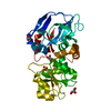
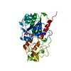
| Structure viewer | Molecule: MolmilJmol/JSmol |
|---|
- Downloads & links
-Download
| PDBx/mmCIF format | 1z8g.cif.gz | 96.4 KB | Display | PDBx/mmCIF format |
|---|---|---|---|---|
| PDB format | pdb1z8g.ent.gz | 70.5 KB | Display | PDB format |
| PDBx/mmJSON format | 1z8g.json.gz | Tree view | PDBx/mmJSON format | |
| Others |  Other downloads Other downloads |
-Validation report
| Arichive directory | https://data.pdbj.org/pub/pdb/validation_reports/z8/1z8gftp://data.pdbj.org/pub/pdb/validation_reports/z8/1z8g | HTTPS FTP |
|---|
-Related structure data
| Related structure data |  1ekbS S: Starting model for refinement |
|---|---|
| Similar structure data |
-Links
 PDBj
PDBj- Assembly
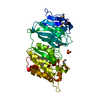
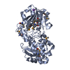
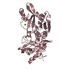
| Deposited unit | 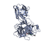
| ||||||||
|---|---|---|---|---|---|---|---|---|---|
| 1 |
| ||||||||
| Unit cell |
|
-Components
| #1: Protein | Mass: 40448.766 Da / Num. of mol.: 1 / Mutation: N112A Source method: isolated from a genetically manipulated source Source: (gene. exp.) Homo sapiens (human) / Gene: HPN, TMPRSS1 / Production host:  Pichia pastoris (fungus) Pichia pastoris (fungus)References: UniProt: P05981, Hydrolases; Acting on peptide bonds (peptidases); Serine endopeptidases |
|---|---|
| #2: Protein/peptide | 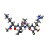  Type: Peptide-like / Class: Inhibitor / Mass: 606.202 Da / Num. of mol.: 1 / Source method: obtained synthetically / Details: THE PEPTIDE WAS CHEMICALLY SYNTHESIZED. Type: Peptide-like / Class: Inhibitor / Mass: 606.202 Da / Num. of mol.: 1 / Source method: obtained synthetically / Details: THE PEPTIDE WAS CHEMICALLY SYNTHESIZED.References: N-acetyl-6-ammonio-L-norleucyl-L-glutaminyl-N-[(1S)-4-{[amino(iminio)methyl]amino}-1-(chloroacetyl)butyl]-L-leucinamide |
| #3: Water | ChemComp-HOH /  Mass: 18.015 Da / Num. of mol.: 461 / Source method: isolated from a natural source / Formula: H2O Mass: 18.015 Da / Num. of mol.: 461 / Source method: isolated from a natural source / Formula: H2O |
| Compound details | THE UNBOUND FORM OF THE INHIBITOR (CHAIN L) IS ACE-LYS-GLN-LEU-ARG-CHLOROMETHYLKETONE. UPON ...THE UNBOUND FORM OF THE INHIBITOR (CHAIN L) IS ACE-LYS-GLN-LEU-ARG-CHLOROMETH |
| Has protein modification | Y |
| Sequence details | THERE IS A CHAIN BREAK BETWEEN RESIDUES 162 AND 163 (RESIDUES 160-162 ARE DISORDERED). THIS PEPTIDE ...THERE IS A CHAIN BREAK BETWEEN RESIDUES 162 AND 163 (RESIDUES 160-162 ARE DISORDERED |
-Experimental details
-Experiment
| Experiment | Method: X-RAY DIFFRACTION / Number of used crystals: 1 |
|---|
- Sample preparation
| Crystal | Density Matthews: 2.16 Å3/Da / Density % sol: 42.5 % |
|---|---|
| Crystal grow | Temperature: 293 K / Method: vapor diffusion, sitting drop / pH: 5.6 Details: PEG 3350, ammonium fluoride, Na-cacodylate, pH 5.6, VAPOR DIFFUSION, SITTING DROP, temperature 293K |
-Data collection
| Diffraction | Mean temperature: 90 K |
|---|---|
| Diffraction source | Source: ROTATING ANODE / Type: RIGAKU RUH3R / Wavelength: 1.5418 Å |
| Detector | Type: RIGAKU RAXIS IV / Detector: IMAGE PLATE / Date: Feb 6, 2004 / Details: OSMIC CONFOCAL OPTIC (GREEN) |
| Radiation | Monochromator: OSMIC MIRRORS / Protocol: SINGLE WAVELENGTH / Monochromatic (M) / Laue (L): M / Scattering type: x-ray |
| Radiation wavelength | Wavelength: 1.5418 Å / Relative weight: 1 |
| Reflection | Resolution: 1.55→23.9 Å / Num. all: 46843 / Num. obs: 46843 / % possible obs: 95 % / Observed criterion σ(F): -3 / Observed criterion σ(I): -3 / Redundancy: 6 % / Biso Wilson estimate: 17 Å2 / Rmerge(I) obs: 0.056 / Net I/σ(I): 21.6 |
| Reflection shell | Resolution: 1.55→1.61 Å / Rmerge(I) obs: 0.327 / Mean I/σ(I) obs: 2.1 / % possible all: 59.2 |
- Processing
| Software |
| ||||||||||||||||||||
|---|---|---|---|---|---|---|---|---|---|---|---|---|---|---|---|---|---|---|---|---|---|
| Refinement | Method to determine structure: MOLECULAR REPLACEMENT Starting model: PDB ENTRY 1EKB Resolution: 1.55→23.9 Å / σ(F): 0 / Stereochemistry target values: Engh & Huber
| ||||||||||||||||||||
| Refinement step | Cycle: LAST / Resolution: 1.55→23.9 Å
| ||||||||||||||||||||
| Refine LS restraints |
|
