 Movie
Movie Controller

[English] 日本語
 Yorodumi
- PDB-1xzq: Structure of the GTP-binding protein TrmE from Thermotoga maritim... -
+ Open data

- Basic information
| Entry | Database: PDB / ID: 1xzq | ||||||
|---|---|---|---|---|---|---|---|
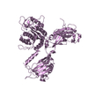
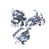
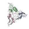
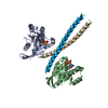
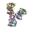
| Title | Structure of the GTP-binding protein TrmE from Thermotoga maritima complexed with 5-formyl-THF | ||||||
 Components Components | (Probable tRNA modification GTPase trmE) x 2 | ||||||
 Keywords Keywords |  HYDROLASE / GTP-binding / THF-binding / tRNA-modification HYDROLASE / GTP-binding / THF-binding / tRNA-modification | ||||||
| Function / homology |  Function and homology information Function and homology informationtRNA wobble uridine modification / tRNA methylation / Hydrolases; Acting on acid anhydrides; Acting on GTP to facilitate cellular and subcellular movement / GTPase activity / GTP binding / metal ion binding / cytosol / cytoplasmSimilarity search - Function | ||||||
| Biological species |   Thermotoga maritima (bacteria) Thermotoga maritima (bacteria) | ||||||
| Method | X-RAY DIFFRACTION / SYNCHROTRON / SAD / Resolution: 2.9 Å | ||||||
 Authors Authors | Scrima, A. / Vetter, I.R. / Armengod, M.E. / Wittinghofer, A. | ||||||
 Citation Citation | Journal: Embo J. / Year: 2005 Title: The structure of the TrmE GTP-binding protein and its implications for tRNA modification Authors: Scrima, A. / Vetter, I.R. / Armengod, M.E. / Wittinghofer, A. | ||||||
| History |
|
- Structure visualization
| Structure viewer | Molecule: MolmilJmol/JSmol |
|---|
- Downloads & links
-Download
| PDBx/mmCIF format | 1xzq.cif.gz | 124.6 KB | Display | PDBx/mmCIF format |
|---|---|---|---|---|
| PDB format | pdb1xzq.ent.gz | 96.7 KB | Display | PDB format |
| PDBx/mmJSON format | 1xzq.json.gz | Tree view | PDBx/mmJSON format | |
| Others |  Other downloads Other downloads |
-Validation report
| Arichive directory | https://data.pdbj.org/pub/pdb/validation_reports/xz/1xzqftp://data.pdbj.org/pub/pdb/validation_reports/xz/1xzq | HTTPS FTP |
|---|
-Related structure data


-Links
 PDBj
PDBj- Assembly
| Deposited unit | 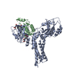
| ||||||||
|---|---|---|---|---|---|---|---|---|---|
| 1 |
| ||||||||
| 2 | 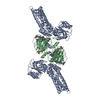
| ||||||||
| Unit cell |
| ||||||||
| Details | The biological assembly is a homodimer (obtained by superimposition of the full-length molecule (A) and the N-terminal domain (B)). Dimerisation via the N-terminal domain is essential for the formation of the THF-binding site. |
-Components
| #1: Protein | Mass: 54189.203 Da / Num. of mol.: 1 Source method: isolated from a genetically manipulated source Source: (gene. exp.) Thermotoga maritima (bacteria) / Gene: TrmE / Plasmid: pET20b / Production host: Escherichia coli (E. coli) / Strain (production host): BL21DE3 (TrmE-) / References: UniProt: Q9WYA4 |
|---|---|
| #2: Protein | Mass: 16363.895 Da / Num. of mol.: 1 / Fragment: N-terminal domain (residues 1-117) Source method: isolated from a genetically manipulated source Source: (gene. exp.) Thermotoga maritima (bacteria) / Gene: TrmE / Plasmid: pET20b / Production host: Escherichia coli (E. coli) / Strain (production host): BL21DE3 (TrmE-) / References: UniProt: Q9WYA4 |
| #3: Chemical |   Mass: 473.439 Da / Num. of mol.: 2 / Source method: obtained synthetically / Formula: C20H23N7O7 Mass: 473.439 Da / Num. of mol.: 2 / Source method: obtained synthetically / Formula: C20H23N7O7 |
-Experimental details
-Experiment
| Experiment | Method: X-RAY DIFFRACTION / Number of used crystals: 1 |
|---|
- Sample preparation
| Crystal | Density Matthews: 4.43 Å3/Da / Density % sol: 72 % |
|---|---|
| Crystal grow | Temperature: 293 K / Method: vapor diffusion, hanging drop / pH: 5.5 Details: ammonium sulphate, mes, pH 5.5, VAPOR DIFFUSION, HANGING DROP, temperature 293K |
-Data collection
| Diffraction | Mean temperature: 100 K |
|---|---|
| Diffraction source | Source: SYNCHROTRON / Site: ESRF  / Beamline: ID14-2 / Wavelength: 0.934 Å / Beamline: ID14-2 / Wavelength: 0.934 Å |
| Radiation | Protocol: SINGLE WAVELENGTH / Monochromatic (M) / Laue (L): M / Scattering type: x-ray |
| Radiation wavelength | Wavelength: 0.934 Å / Relative weight: 1 |
| Reflection | Resolution: 2.9→19.93 Å / Num. all: 24233 / Num. obs: 24138 / % possible obs: 99.6 % / Observed criterion σ(F): 0 / Redundancy: 6.3 % / Biso Wilson estimate: 89.9 Å2 / Rmerge(I) obs: 0.047 / Net I/σ(I): 25.08 |
| Reflection shell | Resolution: 2.9→3 Å / Redundancy: 6.3 % / Rmerge(I) obs: 0.28 / Mean I/σ(I) obs: 7.3 / Num. unique all: 2322 / % possible all: 99.7 |
- Processing
| Software |
| |||||||||||||||||||||||||
|---|---|---|---|---|---|---|---|---|---|---|---|---|---|---|---|---|---|---|---|---|---|---|---|---|---|---|
| Refinement | Method to determine structure: SAD / Resolution: 2.9→19.93 Å / Isotropic thermal model: RESTRAINED / Cross valid method: THROUGHOUT / σ(F): 0 / Stereochemistry target values: Engh & Huber
| |||||||||||||||||||||||||
| Displacement parameters | Biso mean: 77.57 Å2
| |||||||||||||||||||||||||
| Refine analyze |
| |||||||||||||||||||||||||
| Refinement step | Cycle: LAST / Resolution: 2.9→19.93 Å
| |||||||||||||||||||||||||
| Refine LS restraints |
|
