 Movie
Movie Controller

[English] 日本語
 Yorodumi
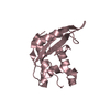
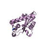
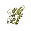
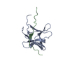
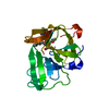
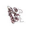
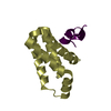
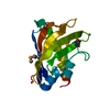
- PDB-1xke: Solution structure of the second Ran-binding domain from human RanBP2 -
+ Open data

- Basic information
| Entry | Database: PDB / ID: 1xke | ||||||
|---|---|---|---|---|---|---|---|
| Title | Solution structure of the second Ran-binding domain from human RanBP2 | ||||||
 Components Components | Ran-binding protein 2 | ||||||
 Keywords Keywords |  PROTEIN TRANSPORT / beta barrel / pleckstrin-homology (PH) domain / phosphotyrosine-binding (PTB) domain PROTEIN TRANSPORT / beta barrel / pleckstrin-homology (PH) domain / phosphotyrosine-binding (PTB) domain | ||||||
| Function / homology |  Function and homology information Function and homology informationcytoplasmic periphery of the nuclear pore complex / SUMO ligase activity / SUMO ligase complex / annulate lamellae / nuclear pore cytoplasmic filaments / Nuclear Pore Complex (NPC) Disassembly / nuclear inclusion body / nuclear pore nuclear basket / Transport of Ribonucleoproteins into the Host Nucleus / Regulation of Glucokinase by Glucokinase Regulatory Protein ...cytoplasmic periphery of the nuclear pore complex / SUMO ligase activity / SUMO ligase complex / annulate lamellae / nuclear pore cytoplasmic filaments / Nuclear Pore Complex (NPC) Disassembly / nuclear inclusion body / nuclear pore nuclear basket / Transport of Ribonucleoproteins into the Host Nucleus / Regulation of Glucokinase by Glucokinase Regulatory Protein / Defective TPR may confer susceptibility towards thyroid papillary carcinoma (TPC) / Transport of the SLBP independent Mature mRNA / Transport of the SLBP Dependant Mature mRNA / NS1 Mediated Effects on Host Pathways / SUMOylation of SUMOylation proteins / Transport of Mature mRNA Derived from an Intronless Transcript / nuclear export / Transferases; Acyltransferases; Aminoacyltransferases / Rev-mediated nuclear export of HIV RNA / Nuclear import of Rev protein / SUMOylation of RNA binding proteins / Transport of Mature mRNA derived from an Intron-Containing Transcript / NEP/NS2 Interacts with the Cellular Export Machinery / tRNA processing in the nucleus / SUMO transferase activity / nucleocytoplasmic transport / centrosome localization / Viral Messenger RNA Synthesis / NLS-bearing protein import into nucleus / regulation of gluconeogenesis / SUMOylation of ubiquitinylation proteins / Vpr-mediated nuclear import of PICs / SUMOylation of DNA replication proteins / protein sumoylation / Signaling by ALK fusions and activated point mutants / Regulation of HSF1-mediated heat shock response / mRNA transport / Amplification of signal from unattached kinetochores via a MAD2 inhibitory signal / SUMOylation of DNA damage response and repair proteins / nuclear pore / Mitotic Prometaphase / EML4 and NUDC in mitotic spindle formation / Resolution of Sister Chromatid Cohesion / response to amphetamine / SUMOylation of chromatin organization proteins / HCMV Late Events / RHO GTPases Activate Formins / Transcriptional regulation by small RNAs / ISG15 antiviral mechanism / small GTPase binding / HCMV Early Events / Separation of Sister Chromatids / protein folding / nuclear envelope / snRNP Assembly / nuclear membrane / intracellular membrane-bounded organelle / protein-containing complex binding / SARS-CoV-2 activates/modulates innate and adaptive immune responses / RNA binding / nucleoplasm / membrane / metal ion binding / cytosol / cytoplasmSimilarity search - Function | ||||||
| Biological species |  Homo sapiens (human) Homo sapiens (human) | ||||||
| Method | SOLUTION NMR / simulated annealing | ||||||
 Authors Authors | Geyer, J.P. / Doeker, R. / Kremer, W. / Zhao, X. / Kuhlmann, J. / Kalbitzer, H.R. | ||||||
 Citation Citation | Journal: J.Mol.Biol. / Year: 2005 Title: Solution structure of the Ran-binding domain 2 of RanBP2 and its interaction with the C terminus of Ran. Authors: Geyer, J.P. / Doker, R. / Kremer, W. / Zhao, X. / Kuhlmann, J. / Kalbitzer, H.R. #1: Journal: J.Biomol.Nmr / Year: 2002 Title: Sequence-specific resonance assignment of the second Ran-binding domain of human RanBP2 Authors: Doeker, R. / Zhao, X. / Kremer, W. / Villa, B. / Kuhlmann, J. / Kalbitzer, H.R. | ||||||
| History |
|
- Structure visualization
| Structure viewer | Molecule: MolmilJmol/JSmol |
|---|
- Downloads & links
-Download
| PDBx/mmCIF format | 1xke.cif.gz | 824.6 KB | Display | PDBx/mmCIF format |
|---|---|---|---|---|
| PDB format | pdb1xke.ent.gz | 717 KB | Display | PDB format |
| PDBx/mmJSON format | 1xke.json.gz | Tree view | PDBx/mmJSON format | |
| Others |  Other downloads Other downloads |
-Validation report
| Arichive directory | https://data.pdbj.org/pub/pdb/validation_reports/xk/1xkeftp://data.pdbj.org/pub/pdb/validation_reports/xk/1xke | HTTPS FTP |
|---|
-Related structure data
| Similar structure data | |
|---|---|
| Other databases |
|


-Links
 PDBj
PDBj


- Assembly
| Deposited unit | 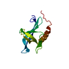
| |||||||||
|---|---|---|---|---|---|---|---|---|---|---|
| 1 |
| |||||||||
| NMR ensembles |
|
-Components
| #1: Protein | Mass: 15138.471 Da / Num. of mol.: 1 / Fragment: Ran-binding domain 2 (RanBD2) Source method: isolated from a genetically manipulated source Source: (gene. exp.) Homo sapiens (human) / Plasmid: pGEX-2T / Species (production host): Escherichia coli / Production host:  Escherichia coli BL21(DE3) (bacteria) / Strain (production host): BL21(DE3) / References: UniProt: P49792 Escherichia coli BL21(DE3) (bacteria) / Strain (production host): BL21(DE3) / References: UniProt: P49792 |
|---|
-Experimental details
-Experiment
| Experiment | Method: SOLUTION NMR | ||||||||||||||||||||
|---|---|---|---|---|---|---|---|---|---|---|---|---|---|---|---|---|---|---|---|---|---|
| NMR experiment |
|
- Sample preparation
| Details |
| |||||||||||||||
|---|---|---|---|---|---|---|---|---|---|---|---|---|---|---|---|---|
| Sample conditions | Ionic strength: 150 mM Na2SO4 / pH: 6.5 / Pressure: ambient / Temperature: 298 K |
-NMR measurement
| Radiation | Protocol: SINGLE WAVELENGTH / Monochromatic (M) / Laue (L): M | |||||||||||||||
|---|---|---|---|---|---|---|---|---|---|---|---|---|---|---|---|---|
| Radiation wavelength | Relative weight: 1 | |||||||||||||||
| NMR spectrometer |
|
- Processing
| NMR software |
| ||||||||||||||||||||||||||||
|---|---|---|---|---|---|---|---|---|---|---|---|---|---|---|---|---|---|---|---|---|---|---|---|---|---|---|---|---|---|
| Refinement | Method: simulated annealing / Software ordinal: 1 Details: The structures are based on a total of 1492 restraints (1281 NOE-derived distance restraints, 182 dihedral angle restraints and 29 hydrogen bond restraints). The structures were also refined ...Details: The structures are based on a total of 1492 restraints (1281 NOE-derived distance restraints, 182 dihedral angle restraints and 29 hydrogen bond restraints). The structures were also refined in water using XPLOR-NIH 2.9.6 using the protocol of Linge et al. (Linge, J. P., Williams, M. A., Spronk, C. A., Bonvin, A. M., & Nilges, M. ,2003. Refinement of protein structures in explicit solvent. Proteins 50, 496-506). | ||||||||||||||||||||||||||||
| NMR representative | Selection criteria: lowest energy | ||||||||||||||||||||||||||||
| NMR ensemble | Conformer selection criteria: structures with the lowest energy Conformers calculated total number: 200 / Conformers submitted total number: 20 |
