 Movie
Movie Controller

[English] 日本語
 Yorodumi
- PDB-3eli: Crystal structure of the AHSA1 (SPO3351) protein from Silicibacte... -
+ Open data

- Basic information
| Entry | Database: PDB / ID: 3eli | ||||||
|---|---|---|---|---|---|---|---|
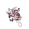
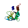
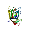
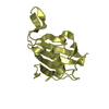
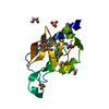
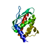
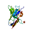
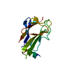
| Title | Crystal structure of the AHSA1 (SPO3351) protein from Silicibacter pomeroyi, Northeast Structural Genomics Consortium Target SiR160 | ||||||
 Components Components | Aha1 domain protein | ||||||
 Keywords Keywords | structural genomics / unknown function / alpha-beta protein / PSI-2 / Protein Structure Initiative / Northeast Structural Genomics Consortium / NESG | ||||||
| Function / homology | Activator of Hsp90 ATPase homologue 1-like / Activator of Hsp90 ATPase homolog 1-like protein / START domain / Alpha-D-Glucose-1,6-Bisphosphate; Chain A, domain 4 / START-like domain superfamily / 2-Layer Sandwich / Alpha Beta / Aha1 domain protein Function and homology information Function and homology information | ||||||
| Biological species |  Silicibacter pomeroyi (bacteria) Silicibacter pomeroyi (bacteria) | ||||||
| Method |  X-RAY DIFFRACTION / SYNCHROTRON / SAD / Resolution: 2.8 Å X-RAY DIFFRACTION / SYNCHROTRON / SAD / Resolution: 2.8 Å | ||||||
 Authors Authors | Forouhar, F. / Su, M. / Seetharaman, J. / Janjua, H. / Xiao, R. / Ciccosanti, C. / Foote, E.L. / Wang, D. / Tong, S. / Everett, J.K. ...Forouhar, F. / Su, M. / Seetharaman, J. / Janjua, H. / Xiao, R. / Ciccosanti, C. / Foote, E.L. / Wang, D. / Tong, S. / Everett, J.K. / Acton, T.B. / Montelione, G.T. / Hunt, J.F. / Tong, L. / Northeast Structural Genomics Consortium (NESG) | ||||||
 Citation Citation | Journal: To be Published Title: Crystal structure of the AHSA1 (SPO3351) protein from Silicibacter pomeroyi, Northeast Structural Genomics Consortium Target SiR160 Authors: Forouhar, F. / Su, M. / Seetharaman, J. / Janjua, H. / Xiao, R. / Ciccosanti, C. / Foote, E.L. / Wang, D. / Tong, S. / Everett, J.K. / Acton, T.B. / Montelione, G.T. / Hunt, J.F. / Tong, L. | ||||||
| History |
|
- Structure visualization
| Structure viewer | Molecule: MolmilJmol/JSmol |
|---|
- Downloads & links
-Download
| PDBx/mmCIF format | 3eli.cif.gz | 41.8 KB | Display | PDBx/mmCIF format |
|---|---|---|---|---|
| PDB format | pdb3eli.ent.gz | 29.2 KB | Display | PDB format |
| PDBx/mmJSON format | 3eli.json.gz | Tree view | PDBx/mmJSON format | |
| Others |  Other downloads Other downloads |
-Validation report
| Arichive directory | https://data.pdbj.org/pub/pdb/validation_reports/el/3eliftp://data.pdbj.org/pub/pdb/validation_reports/el/3eli | HTTPS FTP |
|---|
-Related structure data
| Similar structure data | |
|---|---|
| Other databases |


-Links
 PDBj
PDBj- Assembly
| Deposited unit | 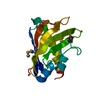
| ||||||||
|---|---|---|---|---|---|---|---|---|---|
| 1 |
| ||||||||
| Unit cell |
|
-Components
| #1: Protein | Mass: 17585.045 Da / Num. of mol.: 1 Source method: isolated from a genetically manipulated source Source: (gene. exp.) Silicibacter pomeroyi (bacteria) / Strain: DSS-3 / Gene: SPO3351 / Plasmid: pET21 / Production host: |
|---|---|
| #2: Water | ChemComp-HOH /  Mass: 18.015 Da / Num. of mol.: 10 / Source method: isolated from a natural source / Formula: H2O Mass: 18.015 Da / Num. of mol.: 10 / Source method: isolated from a natural source / Formula: H2O |
| Has protein modification | Y |
-Experimental details
-Experiment
| Experiment | Method: X-RAY DIFFRACTION / Number of used crystals: 1 |
|---|
- Sample preparation
| Crystal | Density Matthews: 3.51 Å3/Da / Density % sol: 65 % |
|---|---|
| Crystal grow | Method: vapor diffusion, hanging drop / pH: 7.5 Details: Protein solution: 10 mM Tris (pH 7.5), 100 mM sodium chloride, and 5 mM DTT. Reservoir solution: 100 mM TAPS (pH 9) and 8.64 M potassium acetate. VAPOR DIFFUSION, HANGING DROP. |
-Data collection
| Diffraction | Mean temperature: 100 K |
|---|---|
| Diffraction source | Source: SYNCHROTRON / Site: NSLS  / Beamline: X4C / Wavelength: 0.97845 Å / Beamline: X4C / Wavelength: 0.97845 Å |
| Detector | Type: MAR CCD 165 mm / Detector: CCD / Date: Aug 19, 2008 / Details: mirrors |
| Radiation | Monochromator: Si 111 CHANNEL / Protocol: SINGLE WAVELENGTH / Monochromatic (M) / Laue (L): M / Scattering type: x-ray |
| Radiation wavelength | Wavelength: 0.97845 Å / Relative weight: 1 |
| Reflection | Resolution: 2.8→30 Å / Num. all: 11787 / Num. obs: 11776 / % possible obs: 99.9 % / Observed criterion σ(F): 0 / Observed criterion σ(I): 0 / Redundancy: 13.8 % / Biso Wilson estimate: 53 Å2 / Rmerge(I) obs: 0.147 / Rsym value: 0.117 / Net I/σ(I): 21.7 |
| Reflection shell | Resolution: 2.8→2.9 Å / Redundancy: 8.2 % / Rmerge(I) obs: 0.581 / Mean I/σ(I) obs: 2.2 / Num. unique all: 1164 / Rsym value: 0.368 / % possible all: 99.1 |
- Processing
| Software |
| |||||||||||||||||||||||||
|---|---|---|---|---|---|---|---|---|---|---|---|---|---|---|---|---|---|---|---|---|---|---|---|---|---|---|
| Refinement | Method to determine structure: SAD / Resolution: 2.8→19.86 Å / Rfactor Rfree error: 0.012 / Data cutoff high absF: 192372.42 / Data cutoff low absF: 0 / Isotropic thermal model: RESTRAINED / Cross valid method: THROUGHOUT / σ(F): 2 / σ(I): 2 / Stereochemistry target values: Engh & Huber
| |||||||||||||||||||||||||
| Solvent computation | Solvent model: FLAT MODEL / Bsol: 51.6233 Å2 / ksol: 0.4 e/Å3 | |||||||||||||||||||||||||
| Displacement parameters | Biso mean: 63.6 Å2
| |||||||||||||||||||||||||
| Refine analyze |
| |||||||||||||||||||||||||
| Refinement step | Cycle: LAST / Resolution: 2.8→19.86 Å
| |||||||||||||||||||||||||
| Refine LS restraints |
| |||||||||||||||||||||||||
| LS refinement shell | Resolution: 2.8→2.9 Å / Rfactor Rfree error: 0.066 / Total num. of bins used: 10
|
