 Movie
Movie Controller

[English] 日本語
 Yorodumi
- PDB-1x9i: Crystal structure of Crystal structure of phosphoglucose/phosphom... -
+ Open data

- Basic information
| Entry | Database: PDB / ID: 1x9i | ||||||
|---|---|---|---|---|---|---|---|
| Title | Crystal structure of Crystal structure of phosphoglucose/phosphomannose phosphoglucose/phosphomannoseisomerase from Pyrobaculum aerophilum in complex with glucose 6-phosphate | ||||||
 Components Components | glucose-6-phosphate isomerase | ||||||
 Keywords Keywords | ISOMERASE / enzyme / crenarchaeon / hyperthermophile / PGI superfamily / glucose 6-phosphate | ||||||
| Function / homology |  Function and homology informationmannose-6-phosphate isomerase / mannose-6-phosphate isomerase activity / glucose-6-phosphate isomerase / glucose-6-phosphate isomerase activity / carbohydrate derivative metabolic process / carbohydrate derivative binding / carbohydrate metabolic process Function and homology informationmannose-6-phosphate isomerase / mannose-6-phosphate isomerase activity / glucose-6-phosphate isomerase / glucose-6-phosphate isomerase activity / carbohydrate derivative metabolic process / carbohydrate derivative binding / carbohydrate metabolic processSimilarity search - Function | ||||||
| Biological species |   Pyrobaculum aerophilum (archaea) Pyrobaculum aerophilum (archaea) | ||||||
| Method | X-RAY DIFFRACTION / SYNCHROTRON / refinement / Resolution: 1.16 Å | ||||||
 Authors Authors | Swan, M.K. / Hansen, T. / Schoenheit, P. / Davies, C. | ||||||
 Citation Citation | Journal: Biochemistry / Year: 2004 Title: Structural basis for phosphomannose isomerase activity in phosphoglucose isomerase from Pyrobaculum aerophilum: a subtle difference between distantly related enzymes. Authors: Swan, M.K. / Hansen, T. / Schoenheit, P. / Davies, C. #1: Journal: To be publishedTitle: A novel phosphoglucose isomerase/phosphomannose isomerase from the crenarchaeon Pyrobaculum aerophilum is a member of the PGI superfamily: structural evidence at 1.16 A resolution Authors: Swan, M.K. / Hansen, T. / Schoenheit, P. / Davies, C. #2: Journal: Acta Crystallogr.,Sect.D / Year: 2004Title: Crystallization and preliminary X-ray diffraction analysis of phosphoglucose/phosphomannose isomerase from Pyrobaculum aerophilum Authors: Swan, M.K. / Hansen, T. / Schoenheit, P. / Davies, C. | ||||||
| History |
|
- Structure visualization
| Structure viewer | Molecule: MolmilJmol/JSmol |
|---|
- Downloads & links
-Download
| PDBx/mmCIF format | 1x9i.cif.gz | 262.1 KB | Display | PDBx/mmCIF format |
|---|---|---|---|---|
| PDB format | pdb1x9i.ent.gz | 212.6 KB | Display | PDB format |
| PDBx/mmJSON format | 1x9i.json.gz | Tree view | PDBx/mmJSON format | |
| Others |  Other downloads Other downloads |
-Validation report
| Arichive directory | https://data.pdbj.org/pub/pdb/validation_reports/x9/1x9iftp://data.pdbj.org/pub/pdb/validation_reports/x9/1x9i | HTTPS FTP |
|---|
-Related structure data
| Related structure data |  1x9hC  1tzbS S: Starting model for refinement C: citing same article ( |
|---|---|
| Similar structure data |


-Links
 PDBj
PDBj

- Assembly
| Deposited unit | 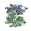
| ||||||||
|---|---|---|---|---|---|---|---|---|---|
| 1 |
| ||||||||
| Unit cell |
| ||||||||
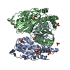
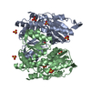
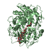
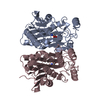
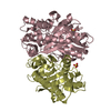
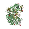
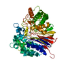
| Details | The biological unit is a dimer. The asymmetric unit is a dimer. |
-Components
| #1: Protein | Mass: 33590.887 Da / Num. of mol.: 2 Source method: isolated from a genetically manipulated source Source: (gene. exp.) Pyrobaculum aerophilum (archaea) / Gene: PAE1610 / Plasmid: pET17b / Production host:  Escherichia coli (E. coli) / Strain (production host): JM109 Escherichia coli (E. coli) / Strain (production host): JM109References: GenBank: 18312750, UniProt: Q8ZWV0*PLUS, glucose-6-phosphate isomerase, mannose-6-phosphate isomerase#2: Chemical | Glucose 6-phosphate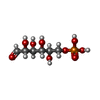  Mass: 260.136 Da / Num. of mol.: 2 Mass: 260.136 Da / Num. of mol.: 2Source method: isolated from a genetically manipulated source Formula: C6H13O9P #3: Chemical | Glycerol  Mass: 92.094 Da / Num. of mol.: 2 / Source method: obtained synthetically / Formula: C3H8O3 Mass: 92.094 Da / Num. of mol.: 2 / Source method: obtained synthetically / Formula: C3H8O3#4: Water | ChemComp-HOH / | Water Mass: 18.015 Da / Num. of mol.: 559 / Source method: isolated from a natural source / Formula: H2O Mass: 18.015 Da / Num. of mol.: 559 / Source method: isolated from a natural source / Formula: H2O |
|---|
-Experimental details
-Experiment
| Experiment | Method: X-RAY DIFFRACTION / Number of used crystals: 1 |
|---|
- Sample preparation
| Crystal | Density Matthews: 2.1 Å3/Da / Density % sol: 42 % |
|---|---|
| Crystal grow | Temperature: 295 K / Method: vapor diffusion, hanging drop / pH: 8.5 Details: 25% polyethylene glycol 8000, 0.22M ammonium sulphate, 100mM Tris, pH 8.5 , VAPOR DIFFUSION, HANGING DROP, temperature 295K |
-Data collection
| Diffraction | Mean temperature: 100 K |
|---|---|
| Diffraction source | Source: SYNCHROTRON / Site: APS  / Beamline: 22-ID / Wavelength: 1 Å / Beamline: 22-ID / Wavelength: 1 Å |
| Detector | Type: MARMOSAIC 225 mm CCD / Detector: CCD / Date: Oct 6, 2003 |
| Radiation | Protocol: SINGLE WAVELENGTH / Monochromatic (M) / Laue (L): M / Scattering type: x-ray |
| Radiation wavelength | Wavelength: 1 Å / Relative weight: 1 |
| Reflection | Resolution: 1.16→36 Å / Num. all: 176109 / Num. obs: 176109 / % possible obs: 90 % / Observed criterion σ(F): 0 / Observed criterion σ(I): 0 / Redundancy: 7.1 % / Biso Wilson estimate: 10 Å2 / Rmerge(I) obs: 0.049 / Rsym value: 0.049 / Net I/σ(I): 40 |
| Reflection shell | Resolution: 1.16→1.2 Å / Redundancy: 4.9 % / Rmerge(I) obs: 0.233 / Mean I/σ(I) obs: 4 / Num. unique all: 15133 / Rsym value: 0.233 / % possible all: 77.5 |
- Processing
| Software |
| ||||||||||||||||||||||||||||||||||||||||||||||||||||||||||||||||||||||||||||||||||||||||||||||||||||||||||||||||||||||||||||||||||||||||||||
|---|---|---|---|---|---|---|---|---|---|---|---|---|---|---|---|---|---|---|---|---|---|---|---|---|---|---|---|---|---|---|---|---|---|---|---|---|---|---|---|---|---|---|---|---|---|---|---|---|---|---|---|---|---|---|---|---|---|---|---|---|---|---|---|---|---|---|---|---|---|---|---|---|---|---|---|---|---|---|---|---|---|---|---|---|---|---|---|---|---|---|---|---|---|---|---|---|---|---|---|---|---|---|---|---|---|---|---|---|---|---|---|---|---|---|---|---|---|---|---|---|---|---|---|---|---|---|---|---|---|---|---|---|---|---|---|---|---|---|---|---|---|
| Refinement | Method to determine structure: refinement Starting model: pdb entry 1tzb Resolution: 1.16→36 Å / Cor.coef. Fo:Fc: 0.973 / Cor.coef. Fo:Fc free: 0.968 / SU B: 0.886 / SU ML: 0.019 / Isotropic thermal model: anisotropic / Cross valid method: THROUGHOUT / σ(F): 0 / ESU R: 0.039 / ESU R Free: 0.036 / Stereochemistry target values: MAXIMUM LIKELIHOOD
| ||||||||||||||||||||||||||||||||||||||||||||||||||||||||||||||||||||||||||||||||||||||||||||||||||||||||||||||||||||||||||||||||||||||||||||
| Solvent computation | Ion probe radii: 0.8 Å / Shrinkage radii: 0.8 Å / VDW probe radii: 1.2 Å / Solvent model: BABINET MODEL WITH MASK | ||||||||||||||||||||||||||||||||||||||||||||||||||||||||||||||||||||||||||||||||||||||||||||||||||||||||||||||||||||||||||||||||||||||||||||
| Displacement parameters | Biso mean: 12.014 Å2
| ||||||||||||||||||||||||||||||||||||||||||||||||||||||||||||||||||||||||||||||||||||||||||||||||||||||||||||||||||||||||||||||||||||||||||||
| Refinement step | Cycle: LAST / Resolution: 1.16→36 Å
| ||||||||||||||||||||||||||||||||||||||||||||||||||||||||||||||||||||||||||||||||||||||||||||||||||||||||||||||||||||||||||||||||||||||||||||
| Refine LS restraints |
| ||||||||||||||||||||||||||||||||||||||||||||||||||||||||||||||||||||||||||||||||||||||||||||||||||||||||||||||||||||||||||||||||||||||||||||
| LS refinement shell | Resolution: 1.16→1.19 Å / Total num. of bins used: 20
|
