 Movie
Movie Controller

[English] 日本語
 Yorodumi
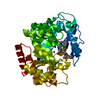
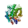
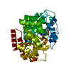
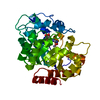
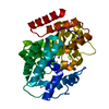
- PDB-1wxy: Crystal structure of adenosine deaminase ligated with a potent in... -
+ Open data

- Basic information
| Entry | Database: PDB / ID: 1wxy | ||||||
|---|---|---|---|---|---|---|---|
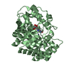
| Title | Crystal structure of adenosine deaminase ligated with a potent inhibitor | ||||||
 Components Components | Adenosine deaminase | ||||||
 Keywords Keywords | HYDROLASE / beta barel | ||||||
| Function / homology |  Function and homology information Function and homology informationnegative regulation of adenosine receptor signaling pathway / inosine biosynthetic process / cytoplasmic vesicle lumen / 2'-deoxyadenosine deaminase activity / adenosine deaminase / adenosine catabolic process / adenosine deaminase activity / hypoxanthine salvage / purine ribonucleoside monophosphate biosynthetic process / nucleotide metabolic process ...negative regulation of adenosine receptor signaling pathway / inosine biosynthetic process / cytoplasmic vesicle lumen / 2'-deoxyadenosine deaminase activity / adenosine deaminase / adenosine catabolic process / adenosine deaminase activity / hypoxanthine salvage / purine ribonucleoside monophosphate biosynthetic process / nucleotide metabolic process / anchoring junction / T cell activation / lysosome / cell adhesion / external side of plasma membrane / zinc ion binding / cytosolSimilarity search - Function | ||||||
| Biological species |  Bos taurus (cattle) Bos taurus (cattle) | ||||||
| Method | X-RAY DIFFRACTION / SYNCHROTRON / MOLECULAR REPLACEMENT / Resolution: 2.5 Å | ||||||
 Authors Authors | Kinoshita, T. | ||||||
 Citation Citation | Journal: Biochemistry / Year: 2005 Title: Structural Basis of Compound Recognition by Adenosine Deaminase Authors: Kinoshita, T. / Nakanishi, I. / Terasaka, T. / Kuno, M. / Seki, N. / Warizaya, M. / Matsumura, H. / Inoue, T. / Takano, K. / Adachi, H. / Mori, Y. / Fujii, T. | ||||||
| History |
|
- Structure visualization
| Structure viewer | Molecule: MolmilJmol/JSmol |
|---|
- Downloads & links
-Download
| PDBx/mmCIF format | 1wxy.cif.gz | 90.2 KB | Display | PDBx/mmCIF format |
|---|---|---|---|---|
| PDB format | pdb1wxy.ent.gz | 68 KB | Display | PDB format |
| PDBx/mmJSON format | 1wxy.json.gz | Tree view | PDBx/mmJSON format | |
| Others |  Other downloads Other downloads |
-Validation report
| Arichive directory | https://data.pdbj.org/pub/pdb/validation_reports/wx/1wxyftp://data.pdbj.org/pub/pdb/validation_reports/wx/1wxy | HTTPS FTP |
|---|
-Related structure data


-Links
 PDBj
PDBj

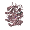
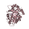
- Assembly
| Deposited unit | 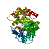
| ||||||||
|---|---|---|---|---|---|---|---|---|---|
| 1 |
| ||||||||
| Unit cell |
|
-Components
| #1: Protein | / Adenosine aminohydrolase Mass: 40341.672 Da / Num. of mol.: 1 / Source method: isolated from a natural source / Source: (natural) Bos taurus (cattle) / Organ: intestine / References: UniProt: P56658, adenosine deaminase |
|---|---|
| #2: Chemical | ChemComp-ZN /   Mass: 65.409 Da / Num. of mol.: 1 / Source method: obtained synthetically / Formula: Zn Mass: 65.409 Da / Num. of mol.: 1 / Source method: obtained synthetically / Formula: Zn |
| #3: Chemical | ChemComp-FRK / 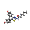  Mass: 382.476 Da / Num. of mol.: 1 / Source method: obtained synthetically / Formula: C21H22N2O3S Mass: 382.476 Da / Num. of mol.: 1 / Source method: obtained synthetically / Formula: C21H22N2O3S |
| #4: Water | ChemComp-HOH / Water Mass: 18.015 Da / Num. of mol.: 251 / Source method: isolated from a natural source / Formula: H2O Mass: 18.015 Da / Num. of mol.: 251 / Source method: isolated from a natural source / Formula: H2O |
-Experimental details
-Experiment
| Experiment | Method: X-RAY DIFFRACTION / Number of used crystals: 1 |
|---|
- Sample preparation
| Crystal | Density Matthews: 2.46 Å3/Da / Density % sol: 49.91 % |
|---|---|
| Crystal grow | Temperature: 293 K / Method: vapor diffusion, sitting drop / pH: 6.5 Details: Ammonium sulphate, PEG400, pH 6.5, VAPOR DIFFUSION, SITTING DROP, temperature 293K |
-Data collection
| Diffraction | Mean temperature: 100 K |
|---|---|
| Diffraction source | Source: SYNCHROTRON / Site: SPring-8  / Beamline: BL24XU / Wavelength: 0.8266 Å / Beamline: BL24XU / Wavelength: 0.8266 Å |
| Detector | Type: RIGAKU RAXIS / Detector: IMAGE PLATE / Date: Dec 1, 2004 |
| Radiation | Monochromator: graphite / Protocol: SINGLE WAVELENGTH / Monochromatic (M) / Laue (L): M / Scattering type: x-ray |
| Radiation wavelength | Wavelength: 0.8266 Å / Relative weight: 1 |
| Reflection | Resolution: 2.5→29.15 Å / Num. all: 14524 / Num. obs: 14422 / % possible obs: 99.3 % / Observed criterion σ(F): 0 / Observed criterion σ(I): 0 / Biso Wilson estimate: 31 Å2 |
- Processing
| Software |
| ||||||||||||||||||||||||||||||||||||||||||||||||||||||||||||||||||||||||||||||||
|---|---|---|---|---|---|---|---|---|---|---|---|---|---|---|---|---|---|---|---|---|---|---|---|---|---|---|---|---|---|---|---|---|---|---|---|---|---|---|---|---|---|---|---|---|---|---|---|---|---|---|---|---|---|---|---|---|---|---|---|---|---|---|---|---|---|---|---|---|---|---|---|---|---|---|---|---|---|---|---|---|---|
| Refinement | Method to determine structure: MOLECULAR REPLACEMENT / Resolution: 2.5→29.15 Å / Rfactor Rfree error: 0.01 / Data cutoff high absF: 1744054.71 / Data cutoff low absF: 0 / Isotropic thermal model: RESTRAINED / Cross valid method: THROUGHOUT / σ(F): 0 / Stereochemistry target values: Engh & Huber
| ||||||||||||||||||||||||||||||||||||||||||||||||||||||||||||||||||||||||||||||||
| Displacement parameters | Biso mean: 38 Å2
| ||||||||||||||||||||||||||||||||||||||||||||||||||||||||||||||||||||||||||||||||
| Refine analyze |
| ||||||||||||||||||||||||||||||||||||||||||||||||||||||||||||||||||||||||||||||||
| Refinement step | Cycle: LAST / Resolution: 2.5→29.15 Å
| ||||||||||||||||||||||||||||||||||||||||||||||||||||||||||||||||||||||||||||||||
| Refine LS restraints |
| ||||||||||||||||||||||||||||||||||||||||||||||||||||||||||||||||||||||||||||||||
| LS refinement shell | Resolution: 2.5→2.66 Å / Rfactor Rfree error: 0.026 / Total num. of bins used: 6
| ||||||||||||||||||||||||||||||||||||||||||||||||||||||||||||||||||||||||||||||||
| Xplor file | Serial no: 1 / Param file: PARMSTD.XPL / Topol file: PROTEIN.TOP |
