- PDB-1vka: Southeast Collaboratory for Structural Genomics: Hypothetical Hum... -
+
Open data
ID or keywords:
Loading...
-
Basic information
Entry
Database: PDB / ID: 1vka
Title
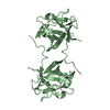
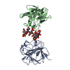
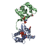
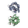
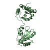
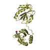
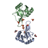
Southeast Collaboratory for Structural Genomics: Hypothetical Human Protein Q15691 N-Terminal Fragment
Components
Microtubule-associated protein RP/EB family member 1
Keywords
Structural Genomics / unknown function / Q15691 / Homo sapiens / PSI / Protein Structure Initiative / Southeast Collaboratory for Structural Genomics / SECSG
Function / homology
Function and homology information
protein localization to astral microtubule / protein localization to mitotic spindle / cortical microtubule cytoskeleton / mitotic spindle astral microtubule end / protein localization to microtubule / microtubule plus-end / cell projection membrane / mitotic spindle microtubule / attachment of mitotic spindle microtubules to kinetochore / microtubule bundle formation ...protein localization to astral microtubule / protein localization to mitotic spindle / cortical microtubule cytoskeleton / mitotic spindle astral microtubule end / protein localization to microtubule / microtubule plus-end / cell projection membrane / mitotic spindle microtubule / attachment of mitotic spindle microtubules to kinetochore / microtubule bundle formation / microtubule plus-end binding / non-motile cilium assembly / protein localization to centrosome / mitotic spindle pole / spindle midzone / negative regulation of microtubule polymerization / microtubule polymerization / microtubule organizing center / establishment of mitotic spindle orientation / regulation of microtubule polymerization or depolymerization / cytoplasmic microtubule / spindle assembly / positive regulation of microtubule polymerization / Amplification of signal from unattached kinetochores via a MAD2 inhibitory signal / Loss of Nlp from mitotic centrosomes / Loss of proteins required for interphase microtubule organization from the centrosome / Recruitment of mitotic centrosome proteins and complexes / Mitotic Prometaphase / Recruitment of NuMA to mitotic centrosomes / Anchoring of the basal body to the plasma membrane / EML4 and NUDC in mitotic spindle formation / protein serine/threonine kinase binding / AURKA Activation by TPX2 / Resolution of Sister Chromatid Cohesion / RHO GTPases Activate Formins / The role of GTSE1 in G2/M progression after G2 checkpoint / Separation of Sister Chromatids / intracellular protein localization / Regulation of PLK1 Activity at G2/M Transition / cell migration / microtubule / ciliary basal body / cadherin binding / cell division / focal adhesion / centrosome / Golgi apparatus / RNA binding / identical protein binding / cytosol Similarity search - Function
SEQUENCE THE STRUCTURE PRESENTED IN THIS ENTRY IS A PROTOLYTIC FRAGMENT (RESIDUES (2-140) OF THE ...SEQUENCE THE STRUCTURE PRESENTED IN THIS ENTRY IS A PROTOLYTIC FRAGMENT (RESIDUES (2-140) OF THE MATURE Q15691 PROTEIN.
Remark 300
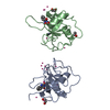
BIOMOLECULE: THIS ENTRY CONTAINS THE CRYSTALLOGRAPHIC ASYMMETRIC UNIT WHICH CONSISTS OF 2 CHAIN(S). ...BIOMOLECULE: THIS ENTRY CONTAINS THE CRYSTALLOGRAPHIC ASYMMETRIC UNIT WHICH CONSISTS OF 2 CHAIN(S). THE BIOLOGICAL UNIT IS UNKNOWN.
Microtubule-associatedproteinRP/EBfamilymember1 / APC-binding protein EB1
Mass: 17395.861 Da / Num. of mol.: 2 / Fragment: N-terminal domain Source method: isolated from a genetically manipulated source Source: (gene. exp.) Homo sapiens (human) Description: The protein was cloned, expressed and purified by the SECSG human protein production group (T.A. Dailey, M. Mayer) under the direction H.A. Dailey. Gene: MAPRE1 / Cell line (production host): BL21(DE53) / Production host: Escherichia coli (E. coli) / Keywords: APC-binding protein, Q15691, sulfur SAS / References: UniProt: Q15691
Method to determine structure: sulfur sas / Resolution: 1.6→100 Å / Cor.coef. Fo:Fc: 0.941 / Cor.coef. Fo:Fc free: 0.936 / SU B: 1.367 / SU ML: 0.05 / SU R Cruickshank DPI: 0.093 / ESU R Free: 0.086 Details: THE Q15691 STRUCTURE WAS INITIALLY SOLVED TO 3 ANGSTROMS IN SPACE GROUP P 21 21 21 USING A SET OF SULFUR SAS DATA COLLECTED WITH FOCUSED CHROMIUM X-RAYS (LAMBDA = 2.29 ANGSTROMS). THE MODEL ...Details: THE Q15691 STRUCTURE WAS INITIALLY SOLVED TO 3 ANGSTROMS IN SPACE GROUP P 21 21 21 USING A SET OF SULFUR SAS DATA COLLECTED WITH FOCUSED CHROMIUM X-RAYS (LAMBDA = 2.29 ANGSTROMS). THE MODEL PRESENTED IN THIS ENTRY WAS REFINED AGAINST 1.6 ANGSTROM DATA COLLECTED AT SER-CAT (22ID) ON A BETTER DIFFRACTING MONOCLINIC CRYSTAL.
Rfactor
Num. reflection
% reflection
Rfree
0.2137
1964
5.004 %
Rwork
0.1998
-
-
all
0.201
-
-
obs
-
37287
-
Solvent computation
Ion probe radii: 0.8 Å / Shrinkage radii: 0.8 Å / VDW probe radii: 1.4 Å / Solvent model: BABINET MODEL PLUS MASK
Displacement parameters
Biso mean: 15.686 Å2
Baniso -1
Baniso -2
Baniso -3
1-
-0.551 Å2
0 Å2
-0.078 Å2
2-
-
0.595 Å2
0 Å2
3-
-
-
-0.05 Å2
Refinement step
Cycle: LAST / Resolution: 1.6→100 Å
Protein
Nucleic acid
Ligand
Solvent
Total
Num. atoms
2070
0
0
201
2271
Refine LS restraints
Refine-ID
Type
Dev ideal
Dev ideal target
Number
X-RAY DIFFRACTION
r_bond_refined_d
0.014
0.022
2120
X-RAY DIFFRACTION
r_angle_refined_deg
1.185
1.931
2866
X-RAY DIFFRACTION
r_dihedral_angle_1_deg
4.871
5
262
X-RAY DIFFRACTION
r_dihedral_angle_3_deg
0.086
0.2
310
X-RAY DIFFRACTION
r_gen_planes_refined
0.006
0.02
1615
X-RAY DIFFRACTION
r_nbd_refined
0.193
0.2
953
X-RAY DIFFRACTION
r_nbtor_refined
0.31
0.2
1535
X-RAY DIFFRACTION
r_xyhbond_nbd_refined
0.119
0.2
125
X-RAY DIFFRACTION
r_symmetry_vdw_refined
0.174
0.2
28
X-RAY DIFFRACTION
r_symmetry_hbond_refined
0.068
0.2
5
X-RAY DIFFRACTION
r_mcbond_it
0.74
1.5
1303
X-RAY DIFFRACTION
r_mcangle_it
1.36
2
2086
X-RAY DIFFRACTION
r_scbond_it
2.135
3
817
X-RAY DIFFRACTION
r_scangle_it
3.337
4.5
780
LS refinement shell
Refine-ID: X-RAY DIFFRACTION / Total num. of bins used: 20
Resolution (Å)
Rfactor Rfree
Num. reflection Rfree
Rfactor Rwork
Num. reflection Rwork
Num. reflection all
1.599-1.6408
0.239
136
0.195
2529
2927
1.641-1.6858
0.222
146
0.186
2715
2926
1.686-1.7346
0.202
146
0.186
2568
2754
1.735-1.788
0.231
112
0.2
2556
2721
1.788-1.8466
0.231
144
0.197
2448
2628
1.847-1.9114
0.223
130
0.196
2393
2556
1.911-1.9835
0.248
120
0.199
2342
2496
1.984-2.0645
0.23
111
0.199
2186
2316
2.064-2.1562
0.215
106
0.202
2159
2287
2.156-2.2614
0.198
105
0.203
2069
2192
2.261-2.3837
0.176
104
0.196
1952
2068
2.384-2.5282
0.193
89
0.199
1858
1956
2.528-2.7026
0.213
102
0.206
1744
1852
2.703-2.919
0.215
87
0.212
1626
1720
2.919-3.1973
0.205
96
0.201
1510
1613
3.197-3.5742
0.24
74
0.204
1340
1436
3.574-4.1263
0.204
60
0.186
1170
1266
4.126-5.0515
0.186
43
0.183
1008
1089
5.052-7.1348
0.228
39
0.217
775
852
7.135-100
0.262
14
0.266
339
486
+
About Yorodumi
-
News
-
Feb 9, 2022. New format data for meta-information of EMDB entries
New format data for meta-information of EMDB entries
Version 3 of the EMDB header file is now the official format.
The previous official version 1.9 will be removed from the archive.
In the structure databanks used in Yorodumi, some data are registered as the other names, "COVID-19 virus" and "2019-nCoV". Here are the details of the virus and the list of structure data.
Jan 31, 2019. EMDB accession codes are about to change! (news from PDBe EMDB page)
EMDB accession codes are about to change! (news from PDBe EMDB page)
The allocation of 4 digits for EMDB accession codes will soon come to an end. Whilst these codes will remain in use, new EMDB accession codes will include an additional digit and will expand incrementally as the available range of codes is exhausted. The current 4-digit format prefixed with “EMD-” (i.e. EMD-XXXX) will advance to a 5-digit format (i.e. EMD-XXXXX), and so on. It is currently estimated that the 4-digit codes will be depleted around Spring 2019, at which point the 5-digit format will come into force.
The EM Navigator/Yorodumi systems omit the EMD- prefix.
Related info.:Q: What is EMD? / ID/Accession-code notation in Yorodumi/EM Navigator
Yorodumi is a browser for structure data from EMDB, PDB, SASBDB, etc.
This page is also the successor to EM Navigator detail page, and also detail information page/front-end page for Omokage search.
The word "yorodu" (or yorozu) is an old Japanese word meaning "ten thousand". "mi" (miru) is to see.
Related info.:EMDB / PDB / SASBDB / Comparison of 3 databanks / Yorodumi Search / Aug 31, 2016. New EM Navigator & Yorodumi / Yorodumi Papers / Jmol/JSmol / Function and homology information / Changes in new EM Navigator and Yorodumi
 Movie
Movie Controller

 Yorodumi
 Open data
 Basic information
 Components
 Keywords
 Function and homology information
 Homo sapiens (human)
 X-RAY DIFFRACTION /
  Authors
 Citation
 Structure visualization
 Downloads & links
 Other downloads
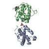
 PDBj
PDBj


 Assembly
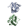

 Mass: 18.015 Da / Num. of mol.: 201 / Source method: isolated from a natural source / Formula: H2O
 Sample preparation
 / Beamline: 22-ID / Wavelength: 1
  Processing