 Movie
Movie Controller

[English] 日本語
 Yorodumi
- PDB-2axm: HEPARIN-LINKED BIOLOGICALLY-ACTIVE DIMER OF FIBROBLAST GROWTH FACTOR -
+ Open data

- Basic information
| Entry | Database: PDB / ID: 2axm | |||||||||
|---|---|---|---|---|---|---|---|---|---|---|
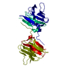
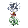
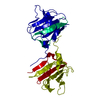
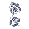
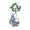
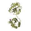
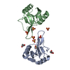
| Title | HEPARIN-LINKED BIOLOGICALLY-ACTIVE DIMER OF FIBROBLAST GROWTH FACTOR | |||||||||
 Components Components | ACIDIC FIBROBLAST GROWTH FACTOR | |||||||||
 Keywords Keywords | GROWTH FACTOR / HUMAN ACIDIC FIBROBLAST GROWTH FACTOR / HEPARIN DECASACCHARIDE / HEXAGONAL CRYSTAL FORM | |||||||||
| Function / homology |  Function and homology information Function and homology informationmesonephric epithelium development / branch elongation involved in ureteric bud branching / regulation of endothelial tube morphogenesis / FGFR3b ligand binding and activation / regulation of endothelial cell chemotaxis to fibroblast growth factor / Signaling by activated point mutants of FGFR3 / FGFR3c ligand binding and activation / Phospholipase C-mediated cascade; FGFR3 / fibroblast growth factor receptor binding / FGFR2b ligand binding and activation ...mesonephric epithelium development / branch elongation involved in ureteric bud branching / regulation of endothelial tube morphogenesis / FGFR3b ligand binding and activation / regulation of endothelial cell chemotaxis to fibroblast growth factor / Signaling by activated point mutants of FGFR3 / FGFR3c ligand binding and activation / Phospholipase C-mediated cascade; FGFR3 / fibroblast growth factor receptor binding / FGFR2b ligand binding and activation / FGFR2c ligand binding and activation / Activated point mutants of FGFR2 / FGFR4 ligand binding and activation / Phospholipase C-mediated cascade; FGFR2 / FGFR1b ligand binding and activation / Phospholipase C-mediated cascade; FGFR4 / organ induction / Signaling by activated point mutants of FGFR1 / FGFR1c ligand binding and activation / Downstream signaling of activated FGFR1 / Phospholipase C-mediated cascade: FGFR1 / S100 protein binding / activation of protein kinase B activity / Signaling by FGFR2 IIIa TM / PI-3K cascade:FGFR3 / PI-3K cascade:FGFR2 / PI-3K cascade:FGFR4 / positive regulation of sprouting angiogenesis / PI-3K cascade:FGFR1 / positive regulation of MAP kinase activity / positive regulation of cell division / positive regulation of intracellular signal transduction / fibroblast growth factor receptor signaling pathway / PI3K Cascade / anatomical structure morphogenesis / SHC-mediated cascade:FGFR3 / SHC-mediated cascade:FGFR2 / SHC-mediated cascade:FGFR4 / SHC-mediated cascade:FGFR1 / FRS-mediated FGFR3 signaling / FRS-mediated FGFR2 signaling / FRS-mediated FGFR4 signaling / FRS-mediated FGFR1 signaling / Signaling by FGFR3 in disease / Signaling by FGFR2 in disease / Signaling by FGFR1 in disease / lung development / positive regulation of endothelial cell migration / epithelial cell proliferation / neurogenesis / regulation of cell migration / positive regulation of epithelial cell proliferation / growth factor activity / wound healing / Negative regulation of FGFR3 signaling / Negative regulation of FGFR2 signaling / Negative regulation of FGFR4 signaling / Negative regulation of FGFR1 signaling / positive regulation of cholesterol biosynthetic process / integrin binding / Constitutive Signaling by Aberrant PI3K in Cancer / positive regulation of angiogenesis / PIP3 activates AKT signaling / heparin binding / cellular response to heat / PI5P, PP2A and IER3 Regulate PI3K/AKT Signaling / extracellular matrix / RAF/MAP kinase cascade / angiogenesis / cell cortex / positive regulation of ERK1 and ERK2 cascade / positive regulation of MAPK cascade / positive regulation of cell migration / positive regulation of cell population proliferation / signal transduction / positive regulation of transcription by RNA polymerase II / : / extracellular region / nucleoplasm / nucleus / cytoplasm / cytosol Similarity search - Function | |||||||||
| Biological species |  Homo sapiens (human) Homo sapiens (human) | |||||||||
| Method |  X-RAY DIFFRACTION / SYNCHROTRON / MOLECULAR REPLACEMENT / Resolution: 3 Å X-RAY DIFFRACTION / SYNCHROTRON / MOLECULAR REPLACEMENT / Resolution: 3 Å | |||||||||
 Authors Authors | Digabriele, A.D. / Lax, I. / Chen, D.I. / Svahn, C.M. / Jaye, M. / Schlessinger, J. / Hendrickson, W.A. | |||||||||
 Citation Citation | Journal: Nature / Year: 1998 Title: Structure of a heparin-linked biologically active dimer of fibroblast growth factor. Authors: DiGabriele, A.D. / Lax, I. / Chen, D.I. / Svahn, C.M. / Jaye, M. / Schlessinger, J. / Hendrickson, W.A. #1: Journal: Biochemistry / Year: 1996Title: X-ray crystal structure of human acidic fibroblast growth factor. Authors: Blaber, M. / DiSalvo, J. / Thomas, K.A. #2: Journal: Science / Year: 1996Title: Heparin Structure and Interactions with Basic Fibroblast Growth Factor Authors: Faham, S. / Hileman, R.E. / Fromm, J.R. / Linhardt, R.J. / Rees, D.C. #3: Journal: Structure / Year: 1993Title: Structural Studies of the Binding of the Anti-Ulcer Drug Sucrose Octasulfate to Acidic Fibroblast Growth Factor Authors: Zhu, X. / Hsu, B.T. / Rees, D.C. | |||||||||
| History |
|
- Structure visualization
| Structure viewer | Molecule: MolmilJmol/JSmol |
|---|
- Downloads & links
-Download
| PDBx/mmCIF format | 2axm.cif.gz | 67.1 KB | Display | PDBx/mmCIF format |
|---|---|---|---|---|
| PDB format | pdb2axm.ent.gz | 50 KB | Display | PDB format |
| PDBx/mmJSON format | 2axm.json.gz | Tree view | PDBx/mmJSON format | |
| Others |  Other downloads Other downloads |
-Validation report
| Arichive directory | https://data.pdbj.org/pub/pdb/validation_reports/ax/2axmftp://data.pdbj.org/pub/pdb/validation_reports/ax/2axm | HTTPS FTP |
|---|
-Related structure data
| Related structure data |  1axmSC S: Starting model for refinement C: citing same article ( |
|---|---|
| Similar structure data |


-Links
 PDBj
PDBj


- Assembly
| Deposited unit | 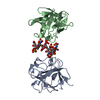
| ||||||||
|---|---|---|---|---|---|---|---|---|---|
| 1 |
| ||||||||
| Unit cell |
| ||||||||
| Noncrystallographic symmetry (NCS) | NCS oper: (Code: given Matrix: (-0.43597, -0.70557, 0.55867), Vector: |
-Components
| #1: Protein | Mass: 15289.199 Da / Num. of mol.: 2 Source method: isolated from a genetically manipulated source Source: (gene. exp.) Homo sapiens (human) / Tissue: NERVE / Cell: ENDOTHELIAL / Cell line: JM109 DE3 / Cellular location: EXTRACELLULAR MATRIX / Gene: ECGF / Organ: BRAIN STEM / Plasmid: PET-3A / Cell line (production host): JM109 DE3 / Cellular location (production host): CYTOPLASM / Production host:  #2: Polysaccharide | 2-O-sulfo-alpha-L-idopyranuronic acid-(1-4)-2-deoxy-6-O-sulfo-2-(sulfoamino)-alpha-D-glucopyranose- ...2-O-sulfo-alpha-L-idopyranuronic acid-(1-4)-2-deoxy-6-O-sulfo-2-(sulfoamino)-alpha-D-glucopyranose-(1-4)-2-O-sulfo-alpha-L-idopyranuronic acid-(1-4)-2-deoxy-6-O-sulfo-2-(sulfoamino)-alpha-D-glucopyranose-(1-4)-2-O-sulfo-alpha-L-idopyranuronic acid-(1-4)-2-deoxy-6-O-sulfo-2-(sulfoamino)-alpha-D-glucopyranose | Source method: isolated from a genetically manipulated source #3: Water | ChemComp-HOH / |  Mass: 18.015 Da / Num. of mol.: 13 / Source method: isolated from a natural source / Formula: H2O Mass: 18.015 Da / Num. of mol.: 13 / Source method: isolated from a natural source / Formula: H2ONonpolymer details | FOR THE DECASACCHA | |
|---|
-Experimental details
-Experiment
| Experiment | Method: X-RAY DIFFRACTION / Number of used crystals: 1 |
|---|
- Sample preparation
| Crystal | Density Matthews: 3.25 Å3/Da / Density % sol: 63 % | |||||||||||||||||||||||||||||||||||
|---|---|---|---|---|---|---|---|---|---|---|---|---|---|---|---|---|---|---|---|---|---|---|---|---|---|---|---|---|---|---|---|---|---|---|---|---|
| Crystal grow | pH: 7 Details: PROTEIN/HEPARIN COMPLEX WAS CRYSTALLIZED FROM 25% PEG 8000, 200 MM MGSO4, 100 MM HEPES, PH 7.0; CRYSTAL WAS SOAKED IN 22% XYLITOL PRIOR TO DATA COLLECTION. | |||||||||||||||||||||||||||||||||||
| Crystal | *PLUS | |||||||||||||||||||||||||||||||||||
| Crystal grow | *PLUS Temperature: 20 ℃ / Method: vapor diffusion, hanging drop | |||||||||||||||||||||||||||||||||||
| Components of the solutions | *PLUS
|
-Data collection
| Diffraction | Mean temperature: 110 K |
|---|---|
| Diffraction source | Source: SYNCHROTRON / Site: NSLS  / Beamline: X4A / Wavelength: 0.98008 / Beamline: X4A / Wavelength: 0.98008 |
| Detector | Type: FUJI / Detector: IMAGE PLATE / Date: Aug 8, 1994 / Details: BENT MIRROR |
| Radiation | Monochromator: SI(111) / Monochromatic (M) / Laue (L): M / Scattering type: x-ray |
| Radiation wavelength | Wavelength: 0.98008 Å / Relative weight: 1 |
| Reflection | Resolution: 3→20 Å / Num. obs: 9682 / % possible obs: 99.7 % / Observed criterion σ(I): 2 / Redundancy: 10.7 % / Rmerge(I) obs: 0.183 / Rsym value: 0.183 / Net I/σ(I): 3.3 |
| Reflection shell | Resolution: 3→3.12 Å / Redundancy: 9.7 % / Rmerge(I) obs: 0.487 / Mean I/σ(I) obs: 1.5 / Rsym value: 0.487 / % possible all: 99.7 |
- Processing
| Software |
| ||||||||||||||||||||||||||||||||||||||||||||||||||||||||||||||||||||||||||||||||
|---|---|---|---|---|---|---|---|---|---|---|---|---|---|---|---|---|---|---|---|---|---|---|---|---|---|---|---|---|---|---|---|---|---|---|---|---|---|---|---|---|---|---|---|---|---|---|---|---|---|---|---|---|---|---|---|---|---|---|---|---|---|---|---|---|---|---|---|---|---|---|---|---|---|---|---|---|---|---|---|---|---|
| Refinement | Method to determine structure: MOLECULAR REPLACEMENT Starting model: PDB ENTRY 1AXM Resolution: 3→13 Å / Rfactor Rfree error: 0.01 / Data cutoff high absF: 100000 / Data cutoff low absF: 1 / Cross valid method: THROUGHOUT / σ(F): 2
| ||||||||||||||||||||||||||||||||||||||||||||||||||||||||||||||||||||||||||||||||
| Displacement parameters | Biso mean: 28.4 Å2 | ||||||||||||||||||||||||||||||||||||||||||||||||||||||||||||||||||||||||||||||||
| Refinement step | Cycle: LAST / Resolution: 3→13 Å
| ||||||||||||||||||||||||||||||||||||||||||||||||||||||||||||||||||||||||||||||||
| Refine LS restraints |
| ||||||||||||||||||||||||||||||||||||||||||||||||||||||||||||||||||||||||||||||||
| Refine LS restraints NCS | NCS model details: RESTRAINTS / Rms dev Biso : 1.25 Å2 / Rms dev position: 0.1 Å / Weight Biso : 2 | ||||||||||||||||||||||||||||||||||||||||||||||||||||||||||||||||||||||||||||||||
| LS refinement shell | Resolution: 3.03→3.19 Å / Rfactor Rfree error: 0.03 / Total num. of bins used: 7
| ||||||||||||||||||||||||||||||||||||||||||||||||||||||||||||||||||||||||||||||||
| Xplor file |
| ||||||||||||||||||||||||||||||||||||||||||||||||||||||||||||||||||||||||||||||||
| Software | *PLUS Name: X-PLOR / Version: 3.1 / Classification: refinement | ||||||||||||||||||||||||||||||||||||||||||||||||||||||||||||||||||||||||||||||||
| Refine LS restraints | *PLUS
|
