 Movie
Movie Controller


+ Open data

- Basic information
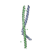
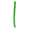
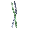
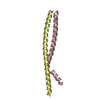
| Entry | Database: PDB / ID: 1uii | ||||||
|---|---|---|---|---|---|---|---|
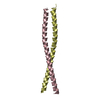
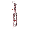
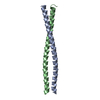
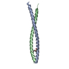
| Title | Crystal structure of Geminin coiled-coil domain | ||||||
 Components Components | Geminin | ||||||
 Keywords Keywords | CELL CYCLE / GEMININ / HUMAN / DNA REPLICATION | ||||||
| Function / homology |  Function and homology information Function and homology informationDNA replication preinitiation complex assembly / Switching of origins to a post-replicative state / negative regulation of DNA-templated DNA replication / positive regulation of chromatin binding / regulation of DNA-templated DNA replication initiation / negative regulation of DNA replication / regulation of DNA replication / negative regulation of cell cycle / Activation of the pre-replicative complex / regulation of mitotic cell cycle ...DNA replication preinitiation complex assembly / Switching of origins to a post-replicative state / negative regulation of DNA-templated DNA replication / positive regulation of chromatin binding / regulation of DNA-templated DNA replication initiation / negative regulation of DNA replication / regulation of DNA replication / negative regulation of cell cycle / Activation of the pre-replicative complex / regulation of mitotic cell cycle / transcription repressor complex / Assembly of the pre-replicative complex / animal organ morphogenesis / histone deacetylase binding / transcription corepressor activity / DNA-binding transcription factor binding / negative regulation of DNA-templated transcription / chromatin binding / nucleoplasm / nucleus / cytosol / cytoplasmSimilarity search - Function | ||||||
| Biological species |  Homo sapiens (human) Homo sapiens (human) | ||||||
| Method | X-RAY DIFFRACTION / SYNCHROTRON / MAD / Resolution: 2 Å | ||||||
 Authors Authors | Yuan, P. / Swaminathan, K. / Robinson, H. | ||||||
 Citation Citation | Journal: Mol.Cell / Year: 2004 Title: A dimerized coiled-coil domain and an adjoining part of geminin interact with two sites on Cdt1 for replication inhibition Authors: Saxena, S. / Yuan, P. / Dhar, S.K. / Senga, T. / Takeda, D. / Robinson, H. / Kornbluth, S. / Swaminathan, K. / Dutta, A. | ||||||
| History |
|
- Structure visualization
| Structure viewer | Molecule: MolmilJmol/JSmol |
|---|
- Downloads & links
-Download
| PDBx/mmCIF format | 1uii.cif.gz | 39.4 KB | Display | PDBx/mmCIF format |
|---|---|---|---|---|
| PDB format | pdb1uii.ent.gz | 27.5 KB | Display | PDB format |
| PDBx/mmJSON format | 1uii.json.gz | Tree view | PDBx/mmJSON format | |
| Others |  Other downloads Other downloads |
-Validation report
| Arichive directory | https://data.pdbj.org/pub/pdb/validation_reports/ui/1uiiftp://data.pdbj.org/pub/pdb/validation_reports/ui/1uii | HTTPS FTP |
|---|
-Related structure data
| Similar structure data |
|---|


-Links
 PDBj
PDBj
- Assembly
| Deposited unit | 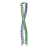
| ||||||||
|---|---|---|---|---|---|---|---|---|---|
| 1 |
| ||||||||
| Unit cell |
|
-Components
| #1: Protein | Mass: 9764.943 Da / Num. of mol.: 2 / Fragment: coiled-coil domain Source method: isolated from a genetically manipulated source Source: (gene. exp.) Homo sapiens (human) / Gene: GMNN / Plasmid: pET28a / Production host:  Escherichia coli (E. coli) / Strain (production host): BL21(DE3)LYS-S / References: UniProt: O75496 Escherichia coli (E. coli) / Strain (production host): BL21(DE3)LYS-S / References: UniProt: O75496#2: Water | ChemComp-HOH / | Water Mass: 18.015 Da / Num. of mol.: 120 / Source method: isolated from a natural source / Formula: H2O Mass: 18.015 Da / Num. of mol.: 120 / Source method: isolated from a natural source / Formula: H2O |
|---|
-Experimental details
-Experiment
| Experiment | Method: X-RAY DIFFRACTION / Number of used crystals: 1 |
|---|
- Sample preparation
| Crystal | Density Matthews: 3.6 Å3/Da / Density % sol: 65.3 % |
|---|---|
| Crystal grow | Temperature: 293 K / Method: evaporation / pH: 8.5 Details: 0.36M Sodium Acetate, 0.1M TRIS, 30% PEG 4000, pH 8.5, EVAPORATION, temperature 293K |
-Data collection
| Diffraction |
| ||||||||||||||||||
|---|---|---|---|---|---|---|---|---|---|---|---|---|---|---|---|---|---|---|---|
| Diffraction source | Source: SYNCHROTRON / Site: NSLS  / Beamline: X12B / Wavelength: 0.9782 / Wavelength: 0.9782, 0.9783, 0.9789, 0.9611 / Beamline: X12B / Wavelength: 0.9782 / Wavelength: 0.9782, 0.9783, 0.9789, 0.9611 | ||||||||||||||||||
| Detector | Type: BRANDEIS / Detector: CCD / Date: Jun 4, 2003 | ||||||||||||||||||
| Radiation |
| ||||||||||||||||||
| Radiation wavelength |
| ||||||||||||||||||
| Reflection | Resolution: 2→30 Å / Num. obs: 25309 / % possible obs: 99.7 % / Redundancy: 6.7 % / Biso Wilson estimate: 13.7 Å2 / Rsym value: 0.085 | ||||||||||||||||||
| Reflection shell | Resolution: 2→2.07 Å / Redundancy: 3.2 % / Rsym value: 0.705 / % possible all: 98.5 |
- Processing
| Software |
| ||||||||||||||||||||||||||||||||||||||||||||||||||||||||||||
|---|---|---|---|---|---|---|---|---|---|---|---|---|---|---|---|---|---|---|---|---|---|---|---|---|---|---|---|---|---|---|---|---|---|---|---|---|---|---|---|---|---|---|---|---|---|---|---|---|---|---|---|---|---|---|---|---|---|---|---|---|---|
| Refinement | Method to determine structure: MAD / Resolution: 2→8 Å / Rfactor Rfree error: 0.005 / Data cutoff high absF: 1162581.49 / Data cutoff low absF: 0 / Isotropic thermal model: RESTRAINED / Cross valid method: THROUGHOUT / σ(F): 3
| ||||||||||||||||||||||||||||||||||||||||||||||||||||||||||||
| Solvent computation | Solvent model: FLAT MODEL / Bsol: 73.4476 Å2 / ksol: 0.577852 e/Å3 | ||||||||||||||||||||||||||||||||||||||||||||||||||||||||||||
| Displacement parameters | Biso mean: 35.6 Å2
| ||||||||||||||||||||||||||||||||||||||||||||||||||||||||||||
| Refine analyze |
| ||||||||||||||||||||||||||||||||||||||||||||||||||||||||||||
| Refinement step | Cycle: LAST / Resolution: 2→8 Å
| ||||||||||||||||||||||||||||||||||||||||||||||||||||||||||||
| Refine LS restraints |
| ||||||||||||||||||||||||||||||||||||||||||||||||||||||||||||
| LS refinement shell | Resolution: 2→2.12 Å / Rfactor Rfree error: 0.017 / Total num. of bins used: 6
| ||||||||||||||||||||||||||||||||||||||||||||||||||||||||||||
| Xplor file |
|
