 Movie
Movie Controller

[English] 日本語
 Yorodumi
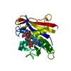
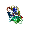
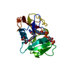
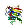
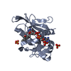
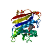
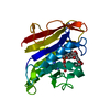
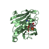
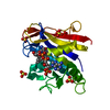
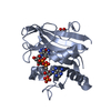
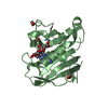
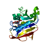
- PDB-1u72: Understanding the Role of Leu22 Variants in Methotrexate Resistan... -
+ Open data

- Basic information
| Entry | Database: PDB / ID: 1u72 | ||||||
|---|---|---|---|---|---|---|---|
| Title | Understanding the Role of Leu22 Variants in Methotrexate Resistance: Comparison of Wild-type and Leu22Arg Variant Mouse and Human Dihydrfolate Reductase Ternary Crystal Complexes with Methotrexate and NADPH | ||||||
 Components Components | Dihydrofolate reductase | ||||||
 Keywords Keywords | OXIDOREDUCTASE / wild type humand DHFR | ||||||
| Function / homology |  Function and homology information Function and homology informationregulation of removal of superoxide radicals / tetrahydrobiopterin biosynthetic process / Metabolism of folate and pterines / tetrahydrofolate metabolic process / sequence-specific mRNA binding / response to methotrexate / dihydrofolate metabolic process / axon regeneration / folic acid binding / G1/S-Specific Transcription ...regulation of removal of superoxide radicals / tetrahydrobiopterin biosynthetic process / Metabolism of folate and pterines / tetrahydrofolate metabolic process / sequence-specific mRNA binding / response to methotrexate / dihydrofolate metabolic process / axon regeneration / folic acid binding / G1/S-Specific Transcription / folic acid metabolic process / glycine biosynthetic process / dihydrofolate reductase / dihydrofolate reductase activity / NADPH binding / Tetrahydrobiopterin (BH4) synthesis, recycling, salvage and regulation / tetrahydrofolate biosynthetic process / mRNA regulatory element binding translation repressor activity / positive regulation of nitric-oxide synthase activity / one-carbon metabolic process / NADP binding / negative regulation of translation / mRNA binding / mitochondrion / cytosolSimilarity search - Function | ||||||
| Biological species |  Homo sapiens (human) Homo sapiens (human) | ||||||
| Method | X-RAY DIFFRACTION / MOLECULAR REPLACEMENT / Resolution: 1.9 Å | ||||||
 Authors Authors | Cody, V. | ||||||
 Citation Citation | Journal: Acta Crystallogr.,Sect.D / Year: 2005 Title: Understanding the role of Leu22 variants in methotrexate resistance: comparison of wild-type and Leu22Arg variant mouse and human dihydrofolate reductase ternary crystal complexes with methotrexate and NADPH. Authors: Cody, V. / Luft, J.R. / Pangborn, W. | ||||||
| History |
|
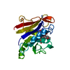
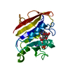
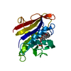
- Structure visualization
| Structure viewer | Molecule: MolmilJmol/JSmol |
|---|
- Downloads & links
-Download
| PDBx/mmCIF format | 1u72.cif.gz | 53.8 KB | Display | PDBx/mmCIF format |
|---|---|---|---|---|
| PDB format | pdb1u72.ent.gz | 38.2 KB | Display | PDB format |
| PDBx/mmJSON format | 1u72.json.gz | Tree view | PDBx/mmJSON format | |
| Others |  Other downloads Other downloads |
-Validation report
| Arichive directory | https://data.pdbj.org/pub/pdb/validation_reports/u7/1u72ftp://data.pdbj.org/pub/pdb/validation_reports/u7/1u72 | HTTPS FTP |
|---|
-Related structure data


-Links
 PDBj
PDBj


- Assembly
| Deposited unit | 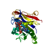
| ||||||||
|---|---|---|---|---|---|---|---|---|---|
| 1 |
| ||||||||
| Unit cell |
|
-Components
| #1: Protein | Mass: 21349.525 Da / Num. of mol.: 1 / Fragment: human DHFR Source method: isolated from a genetically manipulated source Source: (gene. exp.) Homo sapiens (human) / Gene: DHFR / Production host:  Escherichia coli (E. coli) / References: UniProt: P00374, dihydrofolate reductase Escherichia coli (E. coli) / References: UniProt: P00374, dihydrofolate reductase |
|---|---|
| #2: Chemical | ChemComp-NDP / Nicotinamide adenine dinucleotide phosphate  Mass: 745.421 Da / Num. of mol.: 1 / Source method: obtained synthetically / Formula: C21H30N7O17P3 Mass: 745.421 Da / Num. of mol.: 1 / Source method: obtained synthetically / Formula: C21H30N7O17P3 |
| #3: Chemical | ChemComp-MTX / Methotrexate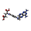  Mass: 454.439 Da / Num. of mol.: 1 / Source method: obtained synthetically / Formula: C20H22N8O5 / Comment: chemotherapy*YM Mass: 454.439 Da / Num. of mol.: 1 / Source method: obtained synthetically / Formula: C20H22N8O5 / Comment: chemotherapy*YM |
| #4: Water | ChemComp-HOH / Water Mass: 18.015 Da / Num. of mol.: 46 / Source method: isolated from a natural source / Formula: H2O Mass: 18.015 Da / Num. of mol.: 46 / Source method: isolated from a natural source / Formula: H2O |
-Experimental details
-Experiment
| Experiment | Method: X-RAY DIFFRACTION / Number of used crystals: 1 |
|---|
- Sample preparation
| Crystal | Density Matthews: 2.64 Å3/Da / Density % sol: 53.41 % |
|---|---|
| Crystal grow | Temperature: 298 K / Method: vapor diffusion, hanging drop / pH: 8 Details: 63% AS, 0.1 M phoshate pH 8.0 , VAPOR DIFFUSION, HANGING DROP, temperature 298K |
-Data collection
| Diffraction | Mean temperature: 298 K |
|---|---|
| Diffraction source | Source: ROTATING ANODE / Type: RIGAKU RU300 / Wavelength: 1.5414 Å |
| Detector | Type: RIGAKU RAXIS IV / Detector: IMAGE PLATE / Date: Feb 6, 1998 / Details: mirrors |
| Radiation | Monochromator: graphite / Protocol: SINGLE WAVELENGTH / Monochromatic (M) / Laue (L): M / Scattering type: x-ray |
| Radiation wavelength | Wavelength: 1.5414 Å / Relative weight: 1 |
| Reflection | Resolution: 1.9→50 Å / Num. all: 8361 / Num. obs: 6408 / % possible obs: 90.3 % / Observed criterion σ(F): 2 / Observed criterion σ(I): 2 / Biso Wilson estimate: 26.5 Å2 / Rmerge(I) obs: 0.049 |
| Reflection shell | Resolution: 1.9→2 Å / % possible all: 55.9 |
- Processing
| Software |
| ||||||||||||||||||||
|---|---|---|---|---|---|---|---|---|---|---|---|---|---|---|---|---|---|---|---|---|---|
| Refinement | Method to determine structure: MOLECULAR REPLACEMENT / Resolution: 1.9→50 Å / σ(F): 2 / Stereochemistry target values: Engh & Huber
| ||||||||||||||||||||
| Refinement step | Cycle: LAST / Resolution: 1.9→50 Å
| ||||||||||||||||||||
| Refine LS restraints |
|
