 Movie
Movie Controller

[English] 日本語
 Yorodumi
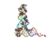
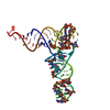
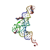
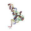
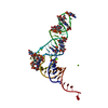
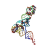
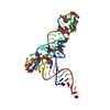
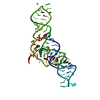
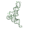
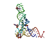
- PDB-1tra: RESTRAINED REFINEMENT OF THE MONOCLINIC FORM OF YEAST PHENYLALANI... -
+ Open data

- Basic information
| Entry | Database: PDB / ID: 1tra | |||||||||
|---|---|---|---|---|---|---|---|---|---|---|
| Title | RESTRAINED REFINEMENT OF THE MONOCLINIC FORM OF YEAST PHENYLALANINE TRANSFER RNA. TEMPERATURE FACTORS AND DYNAMICS, COORDINATED WATERS, AND BASE-PAIR PROPELLER TWIST ANGLES | |||||||||
 Components Components | TRNAPHE | |||||||||
 Keywords Keywords | T-RNA /  SINGLE STRAND / LOOPS SINGLE STRAND / LOOPS | |||||||||
| Function / homology | RNA / RNA (> 10) Function and homology information Function and homology information | |||||||||
| Biological species |  Saccharomyces cerevisiae (brewer's yeast) Saccharomyces cerevisiae (brewer's yeast) | |||||||||
| Method | X-RAY DIFFRACTION / Resolution: 3 Å | |||||||||
 Authors Authors | Westhof, E. / Sundaralingam, M. | |||||||||
 Citation Citation | Journal: Biochemistry / Year: 1986 Title: Restrained refinement of the monoclinic form of yeast phenylalanine transfer RNA. Temperature factors and dynamics, coordinated waters, and base-pair propeller twist angles. Authors: Westhof, E. / Sundaralingam, M. #1: Journal: Acta Crystallogr.,Sect.B / Year: 1978Title: Crystal and Molecular Structure of Yeast Phenylalanyl Transfer RNA. Structure Determination, Difference Fourier Refinement, Molecular Conformation, Metal and Solvent Binding Authors: Stout, C.D. / Mizuno, H. / Rao, S.T. / Swaminathan, P. / Rubin, J. / Brennan, T. / Sundaralingam, M. #2: Journal: Nucleic Acids Res. / Year: 1976Title: Atomic Coordinates and Molecular Conformation of Yeast Phenylalanyl T-RNA, an Independent Investigation Authors: Stout, C.D. / Mizuno, H. / Rubin, J. / Brennan, T. / Rao, S.T. / Sundaralingam, M. #3: Journal: Nucleic Acids Res. / Year: 1976Title: Mechanisms of Chain Folding in Nucleic Acids, the (Omega,Omega) Plot and Its Correlation to the Nucleotide Geometry in Yeast T-RNA(PHE) Authors: Sundaralingam, M. / Mizuno, H. / Stout, C.D. / Rao, S.T. / Liebman, M. / Yathindra, N. #4: Journal: Nature New Biol. / Year: 1972Title: X-Ray Diffraction Study of a New Crystal Form of Yeast Phenylalanine T-RNA Authors: Ichikawa, T. / Sundaralingam, M. | |||||||||
| History |
|
- Structure visualization
| Structure viewer | Molecule: MolmilJmol/JSmol |
|---|
- Downloads & links
-Download
| PDBx/mmCIF format | 1tra.cif.gz | 63.4 KB | Display | PDBx/mmCIF format |
|---|---|---|---|---|
| PDB format | pdb1tra.ent.gz | 43.6 KB | Display | PDB format |
| PDBx/mmJSON format | 1tra.json.gz | Tree view | PDBx/mmJSON format | |
| Others |  Other downloads Other downloads |
-Validation report
| Arichive directory | https://data.pdbj.org/pub/pdb/validation_reports/tr/1traftp://data.pdbj.org/pub/pdb/validation_reports/tr/1tra | HTTPS FTP |
|---|
-Related structure data
| Similar structure data |
|---|
-Links
 PDBj
PDBj


- Assembly
| Deposited unit | 
| ||||||||
|---|---|---|---|---|---|---|---|---|---|
| 1 |
| ||||||||
| Unit cell |
|
-Components
| #1: RNA chain | Mass: 24890.121 Da / Num. of mol.: 1 / Source method: isolated from a natural source / Source: (natural) Saccharomyces cerevisiae (brewer's yeast) | ||
|---|---|---|---|
| #2: Chemical | ChemComp-MG /   Mass: 24.305 Da / Num. of mol.: 5 / Source method: obtained synthetically / Formula: Mg Mass: 24.305 Da / Num. of mol.: 5 / Source method: obtained synthetically / Formula: Mg#3: Water | ChemComp-HOH / | Water Mass: 18.015 Da / Num. of mol.: 110 / Source method: isolated from a natural source / Formula: H2O Mass: 18.015 Da / Num. of mol.: 110 / Source method: isolated from a natural source / Formula: H2O |
-Experimental details
-Experiment
| Experiment | Method: X-RAY DIFFRACTION |
|---|
- Sample preparation
| Crystal | Density Matthews: 2.36 Å3/Da / Density % sol: 47.88 % |
|---|---|
| Crystal grow | *PLUS Method: otherDetails: Ichikawa, T., (1972) Nature New Biol., 236, 174., Ladner, J.E., (1972) J. Mol. Biol., 72, 99. |
-Data collection
| Radiation | Scattering type: x-ray |
|---|---|
| Radiation wavelength | Relative weight: 1 |
| Reflection | *PLUS Highest resolution: 3 Å / Lowest resolution: 10 Å / Num. obs: 4019 / Observed criterion σ(I): 3 |
- Processing
| Software | Name: NUCLSQ / Classification: refinement | |||||||||||||||||||||||||||||||||||||||||||||||||||||||||||||||
|---|---|---|---|---|---|---|---|---|---|---|---|---|---|---|---|---|---|---|---|---|---|---|---|---|---|---|---|---|---|---|---|---|---|---|---|---|---|---|---|---|---|---|---|---|---|---|---|---|---|---|---|---|---|---|---|---|---|---|---|---|---|---|---|---|
| Refinement | Highest resolution: 3 Å /
| |||||||||||||||||||||||||||||||||||||||||||||||||||||||||||||||
| Refinement step | Cycle: LAST / Highest resolution: 3 Å
| |||||||||||||||||||||||||||||||||||||||||||||||||||||||||||||||
| Refine LS restraints |
| |||||||||||||||||||||||||||||||||||||||||||||||||||||||||||||||
| Refinement | *PLUS Highest resolution: 3 Å / Lowest resolution: 10 Å | |||||||||||||||||||||||||||||||||||||||||||||||||||||||||||||||
| Solvent computation | *PLUS | |||||||||||||||||||||||||||||||||||||||||||||||||||||||||||||||
| Displacement parameters | *PLUS |
