 Movie
Movie Controller

[English] 日本語
 Yorodumi
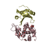
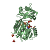
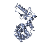
- PDB-1spp: THE CRYSTAL STRUCTURES OF TWO MEMBERS OF THE SPERMADHESIN FAMILY ... -
+ Open data

- Basic information
| Entry | Database: PDB / ID: 1spp | ||||||
|---|---|---|---|---|---|---|---|
| Title | THE CRYSTAL STRUCTURES OF TWO MEMBERS OF THE SPERMADHESIN FAMILY REVEAL THE FOLDING OF THE CUB DOMAIN | ||||||
 Components Components |
| ||||||
 Keywords Keywords | COMPLEX (SEMINAL PLASMA PROTEIN/SPP) / SEMINAL PLASMA PROTEINS / SPERMADHESINS / CUB DOMAIN ARCHITECTURE / COMPLEX (SEMINAL PLASMA PROTEIN-SPP) / COMPLEX (SEMINAL PLASMA PROTEIN-SPP) complex | ||||||
| Function / homology |  Function and homology information Function and homology information | ||||||
| Biological species |  | ||||||
| Method |  X-RAY DIFFRACTION / MIR / Resolution: 2.4 Å X-RAY DIFFRACTION / MIR / Resolution: 2.4 Å | ||||||
 Authors Authors | Romero, A. / Romao, M.J. / Varela, P.F. / Kolln, I. / Dias, J.M. / Carvalho, A.L. / Sanz, L. / Topfer-Petersen, E. / Calvete, J.J. | ||||||
 Citation Citation | Journal: Nat.Struct.Biol. / Year: 1997 Title: The crystal structures of two spermadhesins reveal the CUB domain fold. Authors: Romero, A. / Romao, M.J. / Varela, P.F. / Kolln, I. / Dias, J.M. / Carvalho, A.L. / Sanz, L. / Topfer-Petersen, E. / Calvete, J.J. #1: Journal: FEBS Lett. / Year: 1996Title: Crystallization and Preliminary X-Ray Diffraction Analysis of Boar Seminal Plasma Spermadhesin Psp-I/Psp-II, a Heterodimer of Two Cub Domains Authors: Romero, A. / Varela, P.F. / Sanz, L. / Topfer-Petersen, E. / Calvete, J.J. #2: Journal: FEBS Lett. / Year: 1995Title: Boar Spermadhesin Psp-II: Location of Posttranslational Modifications, Heterodimer Formation with Psp-I Glycoforms and Effect of Dimerization on the Ligand-Binding Capabilities of the Subunits Authors: Calvete, J.J. / Mann, K. / Schafer, W. / Raida, M. / Sanz, L. / Topfer-Petersen, E. #3: Journal: J.Mol.Biol. / Year: 1993Title: The Cub Domain. A Widespread Module in Developmentally Regulated Proteins Authors: Bork, P. / Beckmann, G. | ||||||
| History |
|
- Structure visualization
| Structure viewer | Molecule: MolmilJmol/JSmol |
|---|
- Downloads & links
-Download
| PDBx/mmCIF format | 1spp.cif.gz | 66.8 KB | Display | PDBx/mmCIF format |
|---|---|---|---|---|
| PDB format | pdb1spp.ent.gz | 50.1 KB | Display | PDB format |
| PDBx/mmJSON format | 1spp.json.gz | Tree view | PDBx/mmJSON format | |
| Others |  Other downloads Other downloads |
-Validation report
| Arichive directory | https://data.pdbj.org/pub/pdb/validation_reports/sp/1sppftp://data.pdbj.org/pub/pdb/validation_reports/sp/1spp | HTTPS FTP |
|---|
-Related structure data


-Links
 PDBj
PDBj- Assembly
| Deposited unit | 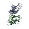
| ||||||||
|---|---|---|---|---|---|---|---|---|---|
| 1 |
| ||||||||
| Unit cell |
|
-Components
| #1: Protein | Mass: 11997.683 Da / Num. of mol.: 1 / Source method: isolated from a natural source / Source: (natural) |
|---|---|
| #2: Protein | Mass: 12658.431 Da / Num. of mol.: 1 / Source method: isolated from a natural source / Source: (natural) |
| #3: Water | ChemComp-HOH /  Mass: 18.015 Da / Num. of mol.: 79 / Source method: isolated from a natural source / Formula: H2O Mass: 18.015 Da / Num. of mol.: 79 / Source method: isolated from a natural source / Formula: H2O |
| Has protein modification | Y |
| Sequence details | ANALYSIS OF CDNAS CODING FOR BOAR SEMINAL PLASMA PSP-I AND PSP-II INDICATED THAT THE FULL-LENGTH ...ANALYSIS OF CDNAS CODING FOR BOAR SEMINAL PLASMA PSP-I AND PSP-II INDICATED THAT THE FULL-LENGTH AMINO ACIDS SEQUENCES OF THE PROTEINS CONTAIN 112 AND 116 RESIDUES, RESPECTIVE |
-Experimental details
-Experiment
| Experiment | Method: X-RAY DIFFRACTION / Number of used crystals: 1 |
|---|
- Sample preparation
| Crystal | Density Matthews: 3.21 Å3/Da / Density % sol: 62 % Description: HEAVY-ATOM DERIVATIVES FOR MIR WERE PREPARED BY USING HEXAGONAL CRYSTALS OF THE PSP-I/PSP-II (CRYSTALS DIFFRACTED UP TO 3.0 A). MIR PHASES WERE CALCULATED USING MLPHARE AFTER SCALING BY ...Description: HEAVY-ATOM DERIVATIVES FOR MIR WERE PREPARED BY USING HEXAGONAL CRYSTALS OF THE PSP-I/PSP-II (CRYSTALS DIFFRACTED UP TO 3.0 A). MIR PHASES WERE CALCULATED USING MLPHARE AFTER SCALING BY SCALEIT. THE MODEL OBTAINED FOR THE HEXAGONAL CRYSTAL FORM WAS USED AS A SEARCH MODEL FOR REFINING THE STRUCTURE IN THE TRIGONAL CRYSTALS TO 2.4 A RESOLUTION BY MOLECULAR REPLACEMENT USING AMORE | |||||||||||||||||||||||||
|---|---|---|---|---|---|---|---|---|---|---|---|---|---|---|---|---|---|---|---|---|---|---|---|---|---|---|
| Crystal grow | pH: 6.5 Details: PROTEIN WAS CRYSTALLIZED FROM 30% (W/V) PEG 2000, 100 MM AMMONIUM ACETATE, PH 6.5. THE INITIAL PROTEIN CONCENTRATION WAS 15 MG/ML | |||||||||||||||||||||||||
| Crystal grow | *PLUS Temperature: 22 ℃ / pH: 7 / Method: vapor diffusion, hanging drop | |||||||||||||||||||||||||
| Components of the solutions | *PLUS
|
-Data collection
| Diffraction | Mean temperature: 285 K |
|---|---|
| Diffraction source | Source: ROTATING ANODE / Type: RIGAKU RUH2R / Wavelength: 1.5418 |
| Detector | Type: MARRESEARCH / Detector: IMAGE PLATE / Date: Nov 1, 1995 |
| Radiation | Monochromator: GRAPHITE(002) / Monochromatic (M) / Laue (L): M / Scattering type: x-ray |
| Radiation wavelength | Wavelength: 1.5418 Å / Relative weight: 1 |
| Reflection | Resolution: 2.4→28.94 Å / Num. obs: 14809 / % possible obs: 97 % / Observed criterion σ(I): 2 / Redundancy: 4.1 % / Biso Wilson estimate: 45.2 Å2 / Rmerge(I) obs: 0.057 / Rsym value: 0.08 / Net I/σ(I): 7.1 |
| Reflection shell | Resolution: 2.4→2.5 Å / Redundancy: 4.1 % / Mean I/σ(I) obs: 3 / Rsym value: 0.265 / % possible all: 98.4 |
| Reflection | *PLUS Num. measured all: 61270 |
| Reflection shell | *PLUS % possible obs: 98.4 % / Rmerge(I) obs: 0.265 |
- Processing
| Software |
| ||||||||||||||||||||||||||||||||||||||||||||||||||||||||||||||||||||||||||||||||
|---|---|---|---|---|---|---|---|---|---|---|---|---|---|---|---|---|---|---|---|---|---|---|---|---|---|---|---|---|---|---|---|---|---|---|---|---|---|---|---|---|---|---|---|---|---|---|---|---|---|---|---|---|---|---|---|---|---|---|---|---|---|---|---|---|---|---|---|---|---|---|---|---|---|---|---|---|---|---|---|---|---|
| Refinement | Method to determine structure: MIR / Resolution: 2.4→8 Å / Data cutoff high absF: 735.54 / Data cutoff low absF: 3.29 / Isotropic thermal model: RESTRAINED / Cross valid method: THROUGHOUT / σ(F): 0
| ||||||||||||||||||||||||||||||||||||||||||||||||||||||||||||||||||||||||||||||||
| Displacement parameters | Biso mean: 31.71 Å2 | ||||||||||||||||||||||||||||||||||||||||||||||||||||||||||||||||||||||||||||||||
| Refinement step | Cycle: LAST / Resolution: 2.4→8 Å
| ||||||||||||||||||||||||||||||||||||||||||||||||||||||||||||||||||||||||||||||||
| Refine LS restraints |
| ||||||||||||||||||||||||||||||||||||||||||||||||||||||||||||||||||||||||||||||||
| LS refinement shell | Resolution: 2.4→2.51 Å / Total num. of bins used: 8
| ||||||||||||||||||||||||||||||||||||||||||||||||||||||||||||||||||||||||||||||||
| Xplor file |
| ||||||||||||||||||||||||||||||||||||||||||||||||||||||||||||||||||||||||||||||||
| Software | *PLUS Name: X-PLOR / Version: 3.1F / Classification: refinement | ||||||||||||||||||||||||||||||||||||||||||||||||||||||||||||||||||||||||||||||||
| Refine LS restraints | *PLUS
|
