 Movie
Movie Controller

[English] 日本語
 Yorodumi
- PDB-1j6r: Crystal structure of Activation (AdoMet binding) domain of Methio... -
+ Open data

- Basic information
| Entry | Database: PDB / ID: 1j6r | ||||||
|---|---|---|---|---|---|---|---|
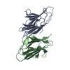
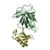
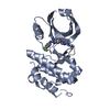
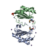
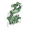
| Title | Crystal structure of Activation (AdoMet binding) domain of Methionine synthase (TM0269) from Thermotoga maritima at 2.2 A resolution | ||||||
 Components Components | METHIONINE SYNTHASE | ||||||
 Keywords Keywords | TRANSFERASE / STRUCTURAL GENOMICS / TM0269 / ACTIVATION (ADOMET BINDING) DOMAIN OF METHIONINE SYNTHASE / JCSG / PSI / Protein Structure Initiative / Joint Center for Structural Genomics | ||||||
| Function / homology | NADH Oxidase - #40 / S-adenosyl-L-methionine dependent methionine synthase, predicted / Vitamin B12-dependent methionine synthase, activation domain superfamily / methionine synthase activity / NADH Oxidase / 3-Layer(aba) Sandwich / Alpha Beta / Methionine synthase Function and homology information Function and homology information | ||||||
| Biological species |   Thermotoga maritima (bacteria) Thermotoga maritima (bacteria) | ||||||
| Method |  X-RAY DIFFRACTION / SYNCHROTRON / MAD / Resolution: 2.3 Å X-RAY DIFFRACTION / SYNCHROTRON / MAD / Resolution: 2.3 Å | ||||||
 Authors Authors | Joint Center for Structural Genomics (JCSG) | ||||||
 Citation Citation | Journal: To be published Title: Crystal structure of Activation (AdoMet binding) domain of Methionine synthase (TM0269) from Thermotoga maritima at 2.2 A resolution Authors: Joint Center for Structural Genomics (JCSG) | ||||||
| History |
|
- Structure visualization
| Structure viewer | Molecule: MolmilJmol/JSmol |
|---|
- Downloads & links
-Download
| PDBx/mmCIF format | 1j6r.cif.gz | 92.1 KB | Display | PDBx/mmCIF format |
|---|---|---|---|---|
| PDB format | pdb1j6r.ent.gz | 70.2 KB | Display | PDB format |
| PDBx/mmJSON format | 1j6r.json.gz | Tree view | PDBx/mmJSON format | |
| Others |  Other downloads Other downloads |
-Validation report
| Arichive directory | https://data.pdbj.org/pub/pdb/validation_reports/j6/1j6rftp://data.pdbj.org/pub/pdb/validation_reports/j6/1j6r | HTTPS FTP |
|---|
-Related structure data
| Similar structure data | |
|---|---|
| Other databases |


-Links
 PDBj
PDBj- Assembly
| Deposited unit | 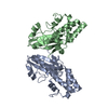
| ||||||||
|---|---|---|---|---|---|---|---|---|---|
| 1 | 
| ||||||||
| 2 | 
| ||||||||
| Unit cell |
|
-Components
| #1: Protein | Mass: 24383.975 Da / Num. of mol.: 2 / Fragment: ACTIVATION (ADOMET BINDING) DOMAIN Source method: isolated from a genetically manipulated source Source: (gene. exp.) Thermotoga maritima (bacteria) / Gene: TM0269 / Production host: #2: Water | ChemComp-HOH / |  Mass: 18.015 Da / Num. of mol.: 103 / Source method: isolated from a natural source / Formula: H2O Mass: 18.015 Da / Num. of mol.: 103 / Source method: isolated from a natural source / Formula: H2O |
|---|
-Experimental details
-Experiment
| Experiment | Method: X-RAY DIFFRACTION / Number of used crystals: 1 |
|---|
- Sample preparation
| Crystal | Density Matthews: 2.99 Å3/Da / Density % sol: 58.81 % |
|---|---|
| Crystal grow | Temperature: 293 K / pH: 7 Details: 2.4 M Ammonium Sulfate; 0.1 M HEPES pH 7.0, VAPOR DIFFUSION, SITTING DROP, NANODROP, temperature 293K, pH 7.00 |
-Data collection
| Diffraction | Mean temperature: 100 K | |||||||||
|---|---|---|---|---|---|---|---|---|---|---|
| Diffraction source | Source: SYNCHROTRON / Site: SSRL  / Beamline: BL9-2 / Wavelength: 0.91837, 0.979029 / Beamline: BL9-2 / Wavelength: 0.91837, 0.979029 | |||||||||
| Detector | Type: ADSC QUANTUM 315 / Detector: CCD / Date: May 12, 2001 | |||||||||
| Radiation | Monochromator: DOUBLE CRYSTAL MONOCHROMATOR / Protocol: MAD / Monochromatic (M) / Laue (L): M / Scattering type: x-ray | |||||||||
| Radiation wavelength |
| |||||||||
| Reflection | Resolution: 2.25→36.22 Å / Num. obs: 27922 / % possible obs: 90.5 % / Redundancy: 4.1 % / Biso Wilson estimate: 43.3 Å2 / Rsym value: 0.048 / Net I/σ(I): 14 | |||||||||
| Reflection shell | Resolution: 2.25→2.37 Å / Redundancy: 3.9 % / Mean I/σ(I) obs: 4.4 / Rsym value: 0.344 / % possible all: 93.5 |
- Processing
| Software |
| ||||||||||||||||||||||||||||||||||||||||||||||||||||||||||||||||||||||||||||||||
|---|---|---|---|---|---|---|---|---|---|---|---|---|---|---|---|---|---|---|---|---|---|---|---|---|---|---|---|---|---|---|---|---|---|---|---|---|---|---|---|---|---|---|---|---|---|---|---|---|---|---|---|---|---|---|---|---|---|---|---|---|---|---|---|---|---|---|---|---|---|---|---|---|---|---|---|---|---|---|---|---|---|
| Refinement | Method to determine structure: MAD / Resolution: 2.3→36.22 Å / Cross valid method: THROUGHOUT / σ(F): 0 Stereochemistry target values: STANDARD CNS DICTIONARY/ENGH AND HUBER Details: DISORDERED SIDECHAINS: CHAIN A: 45, 52,72,76,154,170, 179,180,181,197; CHAIN B: 30,31,34,45,52,72, 76, 86,145,154,163,164,170,178,181,197 DISORDERED RESIDUES: CHAIN A: 177,178 CHAIN B: ...Details: DISORDERED SIDECHAINS: CHAIN A: 45, 52,72,76,154,170, 179,180,181,197; CHAIN B: 30,31,34,45,52,72, 76, 86,145,154,163,164,170,178,181,197 DISORDERED RESIDUES: CHAIN A: 177,178 CHAIN B: 179,180. HYDROGENS HAVE BEEN ADDED IN THE RIDING POSITIONS.
| ||||||||||||||||||||||||||||||||||||||||||||||||||||||||||||||||||||||||||||||||
| Solvent computation | Solvent model: BULK SOLVENT MODELLING / Bsol: 322.79 Å2 / ksol: 0.9 e/Å3 | ||||||||||||||||||||||||||||||||||||||||||||||||||||||||||||||||||||||||||||||||
| Displacement parameters |
| ||||||||||||||||||||||||||||||||||||||||||||||||||||||||||||||||||||||||||||||||
| Refine analyze | Luzzati coordinate error obs: 0.59 Å / Luzzati d res low obs: 6 Å | ||||||||||||||||||||||||||||||||||||||||||||||||||||||||||||||||||||||||||||||||
| Refinement step | Cycle: LAST / Resolution: 2.3→36.22 Å
| ||||||||||||||||||||||||||||||||||||||||||||||||||||||||||||||||||||||||||||||||
| Refine LS restraints |
| ||||||||||||||||||||||||||||||||||||||||||||||||||||||||||||||||||||||||||||||||
| LS refinement shell | Resolution: 2.3→2.41 Å / Total num. of bins used: 20
|
