 Movie
Movie Controller

[English] 日本語
 Yorodumi
- PDB-5v1z: Crystal structure of the RPN13 PRU-RPN2 (932-953)-ubiquitin complex -
+ Open data

- Basic information
| Entry | Database: PDB / ID: 5v1z | |||||||||
|---|---|---|---|---|---|---|---|---|---|---|
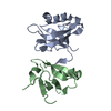
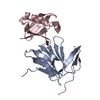
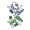
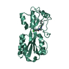
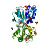
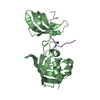
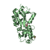
| Title | Crystal structure of the RPN13 PRU-RPN2 (932-953)-ubiquitin complex | |||||||||
 Components Components |
| |||||||||
 Keywords Keywords | PROTEIN BINDING / RPN13 / proteasome / RPN2 / ubiquitin | |||||||||
| Function / homology |  Function and homology information Function and homology informationproteasome accessory complex / proteasome regulatory particle / hypothalamus gonadotrophin-releasing hormone neuron development / female meiosis I / proteasome regulatory particle, lid subcomplex / proteasome regulatory particle, base subcomplex / positive regulation of protein monoubiquitination / fat pad development / seminiferous tubule development / molecular function inhibitor activity ...proteasome accessory complex / proteasome regulatory particle / hypothalamus gonadotrophin-releasing hormone neuron development / female meiosis I / proteasome regulatory particle, lid subcomplex / proteasome regulatory particle, base subcomplex / positive regulation of protein monoubiquitination / fat pad development / seminiferous tubule development / molecular function inhibitor activity / mitochondrion transport along microtubule / cellular response to type I interferon / Regulation of ornithine decarboxylase (ODC) / Proteasome assembly / Cross-presentation of soluble exogenous antigens (endosomes) / Somitogenesis / female gonad development / proteasome binding / regulation of protein catabolic process / male meiosis I / AMPK-induced ERAD and lysosome mediated degradation of PD-L1(CD274) / GSK3B-mediated proteasomal degradation of PD-L1(CD274) / SPOP-mediated proteasomal degradation of PD-L1(CD274) / Ribosome Quality Control (RQC) complex extracts and degrades nascent peptide / proteasome storage granule / positive regulation of intrinsic apoptotic signaling pathway by p53 class mediator / endopeptidase activator activity / energy homeostasis / proteasome assembly / enzyme regulator activity / neuron projection morphogenesis / Maturation of protein E / Maturation of protein E / ER Quality Control Compartment (ERQC) / Myoclonic epilepsy of Lafora / FLT3 signaling by CBL mutants / IRAK2 mediated activation of TAK1 complex / Alpha-protein kinase 1 signaling pathway / Glycogen synthesis / IRAK1 recruits IKK complex / IRAK1 recruits IKK complex upon TLR7/8 or 9 stimulation / Prevention of phagosomal-lysosomal fusion / Endosomal Sorting Complex Required For Transport (ESCRT) / Membrane binding and targetting of GAG proteins / Regulation of TBK1, IKKε (IKBKE)-mediated activation of IRF3, IRF7 / Negative regulation of FLT3 / PTK6 Regulates RTKs and Their Effectors AKT1 and DOK1 / Regulation of TBK1, IKKε-mediated activation of IRF3, IRF7 upon TLR3 ligation / IRAK2 mediated activation of TAK1 complex upon TLR7/8 or 9 stimulation / Constitutive Signaling by NOTCH1 HD Domain Mutants / NOTCH2 Activation and Transmission of Signal to the Nucleus / TICAM1,TRAF6-dependent induction of TAK1 complex / regulation of proteasomal protein catabolic process / TICAM1-dependent activation of IRF3/IRF7 / APC/C:Cdc20 mediated degradation of Cyclin B / regulation of neuron apoptotic process / Downregulation of ERBB4 signaling / APC-Cdc20 mediated degradation of Nek2A / Regulation of FZD by ubiquitination / p75NTR recruits signalling complexes / proteasome complex / InlA-mediated entry of Listeria monocytogenes into host cells / TRAF6 mediated IRF7 activation in TLR7/8 or 9 signaling / NF-kB is activated and signals survival / TRAF6-mediated induction of TAK1 complex within TLR4 complex / Regulation of pyruvate metabolism / positive regulation of protein ubiquitination / Pexophagy / Downregulation of ERBB2:ERBB3 signaling / Regulation of innate immune responses to cytosolic DNA / NRIF signals cell death from the nucleus / Regulation of PTEN localization / regulation of mitochondrial membrane potential / VLDLR internalisation and degradation / Activated NOTCH1 Transmits Signal to the Nucleus / Synthesis of active ubiquitin: roles of E1 and E2 enzymes / Translesion synthesis by REV1 / TICAM1, RIP1-mediated IKK complex recruitment / Regulation of BACH1 activity / Translesion synthesis by POLK / JNK (c-Jun kinases) phosphorylation and activation mediated by activated human TAK1 / InlB-mediated entry of Listeria monocytogenes into host cell / MAP3K8 (TPL2)-dependent MAPK1/3 activation / Activation of IRF3, IRF7 mediated by TBK1, IKKε (IKBKE) / Downregulation of TGF-beta receptor signaling / Translesion synthesis by POLI / Josephin domain DUBs / Gap-filling DNA repair synthesis and ligation in GG-NER / IKK complex recruitment mediated by RIP1 / PINK1-PRKN Mediated Mitophagy / TGF-beta receptor signaling in EMT (epithelial to mesenchymal transition) / TNFR1-induced NF-kappa-B signaling pathway / proteasomal protein catabolic process / Regulation of activated PAK-2p34 by proteasome mediated degradation / TCF dependent signaling in response to WNT / Regulation of NF-kappa B signaling / activated TAK1 mediates p38 MAPK activation / Autodegradation of Cdh1 by Cdh1:APC/C / APC/C:Cdc20 mediated degradation of Securin / NOTCH3 Activation and Transmission of Signal to the Nucleus Similarity search - Function | |||||||||
| Biological species |  Homo sapiens (human) Homo sapiens (human) | |||||||||
| Method |  X-RAY DIFFRACTION / SYNCHROTRON / MOLECULAR REPLACEMENT / Resolution: 2 Å X-RAY DIFFRACTION / SYNCHROTRON / MOLECULAR REPLACEMENT / Resolution: 2 Å | |||||||||
 Authors Authors | Hemmis, C.W. / VanderLinden, R.T. / Yao, T. / Robinson, H. / Hill, C.P. | |||||||||
| Funding support |  United States, 2items United States, 2items
| |||||||||
 Citation Citation | Journal: J. Biol. Chem. / Year: 2017 Title: Structure and energetics of pairwise interactions between proteasome subunits RPN2, RPN13, and ubiquitin clarify a substrate recruitment mechanism. Authors: VanderLinden, R.T. / Hemmis, C.W. / Yao, T. / Robinson, H. / Hill, C.P. | |||||||||
| History |
|
- Structure visualization
| Structure viewer | Molecule: MolmilJmol/JSmol |
|---|
- Downloads & links
-Download
| PDBx/mmCIF format | 5v1z.cif.gz | 96 KB | Display | PDBx/mmCIF format |
|---|---|---|---|---|
| PDB format | pdb5v1z.ent.gz | 72 KB | Display | PDB format |
| PDBx/mmJSON format | 5v1z.json.gz | Tree view | PDBx/mmJSON format | |
| Others |  Other downloads Other downloads |
-Validation report
| Arichive directory | https://data.pdbj.org/pub/pdb/validation_reports/v1/5v1zftp://data.pdbj.org/pub/pdb/validation_reports/v1/5v1z | HTTPS FTP |
|---|
-Related structure data
| Related structure data |  5v1yC 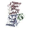 1cmxS  2r2yS C: citing same article ( S: Starting model for refinement |
|---|---|
| Similar structure data |


-Links
 PDBj
PDBj


- Assembly
| Deposited unit | 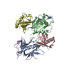
| ||||||||
|---|---|---|---|---|---|---|---|---|---|
| 1 | 
| ||||||||
| 2 | 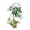
| ||||||||
| Unit cell |
|
-Components
| #1: Protein | Mass: 13482.367 Da / Num. of mol.: 2 / Fragment: PRU domain (UNP residues 19-132) Source method: isolated from a genetically manipulated source Source: (gene. exp.) Homo sapiens (human) / Gene: ADRM1, GP110 / Plasmid: pET151 / Production host:  #2: Protein | Mass: 8576.831 Da / Num. of mol.: 2 Source method: isolated from a genetically manipulated source Source: (gene. exp.) Homo sapiens (human) / Gene: UBB / Plasmid: pET3a / Production host: #3: Protein/peptide | Mass: 2617.699 Da / Num. of mol.: 2 / Fragment: C-termimal domain (UNP residues 932-953) Source method: isolated from a genetically manipulated source Source: (gene. exp.) Homo sapiens (human) / Gene: PSMD1 / Plasmid: pET151 / Production host: #4: Water | ChemComp-HOH / |  Mass: 18.015 Da / Num. of mol.: 81 / Source method: isolated from a natural source / Formula: H2O Mass: 18.015 Da / Num. of mol.: 81 / Source method: isolated from a natural source / Formula: H2O |
|---|
-Experimental details
-Experiment
| Experiment | Method: X-RAY DIFFRACTION / Number of used crystals: 1 |
|---|
- Sample preparation
| Crystal | Density Matthews: 2.24 Å3/Da / Density % sol: 44.97 % |
|---|---|
| Crystal grow | Temperature: 293 K / Method: vapor diffusion, sitting drop / pH: 4.6 / Details: 0.1 M sodium acetate, pH 4.6, 22.5% PEG3350 |
-Data collection
| Diffraction | Mean temperature: 100 K |
|---|---|
| Diffraction source | Source: SYNCHROTRON / Site: SSRL / Beamline: BL7-1 / Wavelength: 1.127 Å |
| Detector | Type: ADSC QUANTUM 315r / Detector: CCD / Date: Feb 17, 2012 |
| Radiation | Monochromator: Si(111) / Protocol: SINGLE WAVELENGTH / Monochromatic (M) / Laue (L): M / Scattering type: x-ray |
| Radiation wavelength | Wavelength: 1.127 Å / Relative weight: 1 |
| Reflection | Resolution: 2→35 Å / Num. obs: 28875 / % possible obs: 100 % / Redundancy: 4.7 % / Rsym value: 0.098 / Net I/σ(I): 16 |
| Reflection shell | Resolution: 2→2.03 Å / Redundancy: 4.6 % / Mean I/σ(I) obs: 2.5 / Num. unique all: 2748 / Num. unique obs: 2748 / Rsym value: 0.628 / % possible all: 100 |
- Processing
| Software |
| |||||||||||||||||||||||||||||||||||||||||||||||||||||||||||||||||||||||||||||
|---|---|---|---|---|---|---|---|---|---|---|---|---|---|---|---|---|---|---|---|---|---|---|---|---|---|---|---|---|---|---|---|---|---|---|---|---|---|---|---|---|---|---|---|---|---|---|---|---|---|---|---|---|---|---|---|---|---|---|---|---|---|---|---|---|---|---|---|---|---|---|---|---|---|---|---|---|---|---|
| Refinement | Method to determine structure: MOLECULAR REPLACEMENT Starting model: PDB entries 2R2Y & 1CMX Resolution: 2→33.012 Å / Cross valid method: FREE R-VALUE / σ(F): 1.98 / Phase error: 21.49
| |||||||||||||||||||||||||||||||||||||||||||||||||||||||||||||||||||||||||||||
| Solvent computation | Shrinkage radii: 0.9 Å / VDW probe radii: 1.11 Å | |||||||||||||||||||||||||||||||||||||||||||||||||||||||||||||||||||||||||||||
| Refinement step | Cycle: LAST / Resolution: 2→33.012 Å
| |||||||||||||||||||||||||||||||||||||||||||||||||||||||||||||||||||||||||||||
| Refine LS restraints |
| |||||||||||||||||||||||||||||||||||||||||||||||||||||||||||||||||||||||||||||
| LS refinement shell |
|
