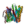
HYDROLASE / Serial crystallography / experimental phasing / in meso crystallization / in situ diffraction data collection / membrane protein structure. / MEMBRANE PROTEIN
Function / homology
Function and homology information
phosphatidylglycerophosphatase / phosphatidylglycerophosphatase activity / phospholipid biosynthetic process / plasma membrane Similarity search - Function
Protocol: SINGLE WAVELENGTH / Monochromatic (M) / Laue (L): M / Scattering type: x-ray
Radiation wavelength
Wavelength: 1.21371 Å / Relative weight: 1
Reflection
Resolution: 1.79→35.614 Å / Num. obs: 39988 / % possible obs: 91.7 % / Redundancy: 2.6 % / CC1/2: 0.92 / Rrim(I) all: 0.1 / Net I/σ(I): 7.93
Reflection shell
Resolution: 1.79→1.9 Å / Redundancy: 2.5 % / Mean I/σ(I) obs: 0.91 / Num. unique obs: 6925 / CC1/2: 0.5 / Rrim(I) all: 1.32 / % possible all: 90
-
Processing
Software
Name
Version
Classification
PHENIX
(1.13_2998: ???)
refinement
XDS
datareduction
XSCALE
datascaling
SHELXCD
phasing
SHELXDE
phasing
Refinement
Method to determine structure: SAD / Resolution: 1.79→35.614 Å / SU ML: 0.27 / Cross valid method: THROUGHOUT / σ(F): 0.85 / Phase error: 31 / Stereochemistry target values: ML
Rfactor
Num. reflection
% reflection
Rfree
0.2624
2168
4.99 %
Rwork
0.2366
-
-
obs
0.2379
39988
90.91 %
Solvent computation
Shrinkage radii: 0.9 Å / VDW probe radii: 1.11 Å / Solvent model: FLAT BULK SOLVENT MODEL
Refinement step
Cycle: LAST / Resolution: 1.79→35.614 Å
Protein
Nucleic acid
Ligand
Solvent
Total
Num. atoms
1579
0
124
73
1776
Refine LS restraints
Refine-ID
Type
Dev ideal
Number
X-RAY DIFFRACTION
f_bond_d
0.005
1754
X-RAY DIFFRACTION
f_angle_d
0.822
2349
X-RAY DIFFRACTION
f_dihedral_angle_d
8.608
1360
X-RAY DIFFRACTION
f_chiral_restr
0.046
266
X-RAY DIFFRACTION
f_plane_restr
0.006
280
LS refinement shell
Resolution (Å)
Rfactor Rfree
Num. reflection Rfree
Rfactor Rwork
Num. reflection Rwork
Refine-ID
% reflection obs (%)
1.7893-1.8309
0.4352
130
0.4147
2544
X-RAY DIFFRACTION
84
1.8309-1.8767
0.365
152
0.3638
2849
X-RAY DIFFRACTION
95
1.8767-1.9275
0.5459
130
0.5361
2431
X-RAY DIFFRACTION
81
1.9275-1.9842
0.367
155
0.3188
2921
X-RAY DIFFRACTION
96
1.9842-2.0482
0.3085
158
0.2953
2858
X-RAY DIFFRACTION
96
2.0482-2.1214
0.3009
149
0.2656
2896
X-RAY DIFFRACTION
96
2.1214-2.2063
0.2884
155
0.2504
2862
X-RAY DIFFRACTION
95
2.2063-2.3067
0.3084
134
0.3175
2666
X-RAY DIFFRACTION
87
2.3067-2.4283
0.2999
144
0.2254
2802
X-RAY DIFFRACTION
94
2.4283-2.5804
0.2218
148
0.2063
2842
X-RAY DIFFRACTION
94
2.5804-2.7796
0.2096
153
0.2041
2803
X-RAY DIFFRACTION
93
2.7796-3.0593
0.2119
143
0.1913
2777
X-RAY DIFFRACTION
92
3.0593-3.5017
0.2295
142
0.199
2704
X-RAY DIFFRACTION
89
3.5017-4.4107
0.2151
137
0.2042
2646
X-RAY DIFFRACTION
87
4.4107-38.4093
0.2794
138
0.2311
2635
X-RAY DIFFRACTION
86
+
About Yorodumi
-
News
-
Feb 9, 2022. New format data for meta-information of EMDB entries
New format data for meta-information of EMDB entries
Version 3 of the EMDB header file is now the official format.
The previous official version 1.9 will be removed from the archive.
In the structure databanks used in Yorodumi, some data are registered as the other names, "COVID-19 virus" and "2019-nCoV". Here are the details of the virus and the list of structure data.
Jan 31, 2019. EMDB accession codes are about to change! (news from PDBe EMDB page)
EMDB accession codes are about to change! (news from PDBe EMDB page)
The allocation of 4 digits for EMDB accession codes will soon come to an end. Whilst these codes will remain in use, new EMDB accession codes will include an additional digit and will expand incrementally as the available range of codes is exhausted. The current 4-digit format prefixed with “EMD-” (i.e. EMD-XXXX) will advance to a 5-digit format (i.e. EMD-XXXXX), and so on. It is currently estimated that the 4-digit codes will be depleted around Spring 2019, at which point the 5-digit format will come into force.
The EM Navigator/Yorodumi systems omit the EMD- prefix.
Related info.:Q: What is EMD? / ID/Accession-code notation in Yorodumi/EM Navigator
Yorodumi is a browser for structure data from EMDB, PDB, SASBDB, etc.
This page is also the successor to EM Navigator detail page, and also detail information page/front-end page for Omokage search.
The word "yorodu" (or yorozu) is an old Japanese word meaning "ten thousand". "mi" (miru) is to see.
Related info.:EMDB / PDB / SASBDB / Comparison of 3 databanks / Yorodumi Search / Aug 31, 2016. New EM Navigator & Yorodumi / Yorodumi Papers / Jmol/JSmol / Function and homology information / Changes in new EM Navigator and Yorodumi
 Movie
Movie Controller


 Open data
 Basic information
 Components
 Keywords
 Function and homology information
 X-RAY DIFFRACTION /
  Authors
 Ireland,
  Switzerland, 2items
  Citation
 Structure visualization
 Downloads & links
 Other downloads
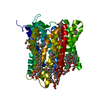


 PDBj
PDBj


 Assembly

 Mass: 356.540 Da / Num. of mol.: 5 / Source method: obtained synthetically / Formula: C21H40O4
 Mass: 247.838 Da / Num. of mol.: 1 / Source method: obtained synthetically / Formula: WO4
 Mass: 18.015 Da / Num. of mol.: 73 / Source method: isolated from a natural source / Formula: H2O
 Sample preparation
 Processing