 Movie
Movie Controller

[English] 日本語
 Yorodumi
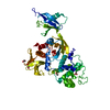
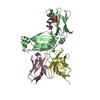
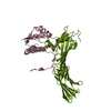
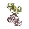
- PDB-1smp: CRYSTAL STRUCTURE OF A COMPLEX BETWEEN SERRATIA MARCESCENS METALL... -
+ Open data

- Basic information
| Entry | Database: PDB / ID: 1smp | ||||||
|---|---|---|---|---|---|---|---|
| Title | CRYSTAL STRUCTURE OF A COMPLEX BETWEEN SERRATIA MARCESCENS METALLO-PROTEASE AND AN INHIBITOR FROM ERWINIA CHRYSANTHEMI | ||||||
 Components Components |
| ||||||
 Keywords Keywords | COMPLEX (METALLOPROTEASE/INHIBITOR) / COMPLEX (METALLOPROTEASE-INHIBITOR) / COMPLEX (METALLOPROTEASE-INHIBITOR) complex | ||||||
| Function / homology |  Function and homology information Function and homology information serralysin / symbiont-mediated killing of host cell / metalloendopeptidase inhibitor activity / extracellular matrix / metalloendopeptidase activity / toxin activity / periplasmic space / calcium ion binding / proteolysis / extracellular space ...serralysin / symbiont-mediated killing of host cell / metalloendopeptidase inhibitor activity / extracellular matrix / metalloendopeptidase activity / toxin activity / periplasmic space / calcium ion binding / proteolysis / extracellular space / zinc ion binding / extracellular region serralysin / symbiont-mediated killing of host cell / metalloendopeptidase inhibitor activity / extracellular matrix / metalloendopeptidase activity / toxin activity / periplasmic space / calcium ion binding / proteolysis / extracellular space ...serralysin / symbiont-mediated killing of host cell / metalloendopeptidase inhibitor activity / extracellular matrix / metalloendopeptidase activity / toxin activity / periplasmic space / calcium ion binding / proteolysis / extracellular space / zinc ion binding / extracellular regionSimilarity search - Function | ||||||
| Biological species |  Serratia marcescens (bacteria)Erwinia chrysanthemi (bacteria) Serratia marcescens (bacteria)Erwinia chrysanthemi (bacteria) | ||||||
| Method | X-RAY DIFFRACTION / Resolution: 2.3 Å | ||||||
 Authors Authors | Baumann, U. / Bauer, M. / Letoffe, S. / Delepelaire, P. / Wandersman, C. | ||||||
 Citation Citation | Journal: J.Mol.Biol. / Year: 1995 Title: Crystal structure of a complex between Serratia marcescens metallo-protease and an inhibitor from Erwinia chrysanthemi. Authors: Baumann, U. / Bauer, M. / Letoffe, S. / Delepelaire, P. / Wandersman, C. #1: Journal: J.Mol.Biol. / Year: 1994Title: Crystal Structure of the 50 kDa Metallo-Proteinase from Serratia Marcescens Authors: Baumann, U. | ||||||
| History |
|
- Structure visualization
| Structure viewer | Molecule: MolmilJmol/JSmol |
|---|
- Downloads & links
-Download
| PDBx/mmCIF format | 1smp.cif.gz | 122 KB | Display | PDBx/mmCIF format |
|---|---|---|---|---|
| PDB format | pdb1smp.ent.gz | 98.9 KB | Display | PDB format |
| PDBx/mmJSON format | 1smp.json.gz | Tree view | PDBx/mmJSON format | |
| Others |  Other downloads Other downloads |
-Validation report
| Arichive directory | https://data.pdbj.org/pub/pdb/validation_reports/sm/1smpftp://data.pdbj.org/pub/pdb/validation_reports/sm/1smp | HTTPS FTP |
|---|
-Related structure data
| Similar structure data |
|---|


-Links
 PDBj
PDBj


- Assembly
| Deposited unit | 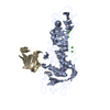
| ||||||||
|---|---|---|---|---|---|---|---|---|---|
| 1 |
| ||||||||
| Unit cell |
|
-Components
| #1: Protein | Mass: 50439.195 Da / Num. of mol.: 1 Source method: isolated from a genetically manipulated source Source: (gene. exp.) Serratia marcescens (bacteria) / Gene: INH / Plasmid: PUC / Gene (production host): INH / Production host: Escherichia coli (E. coli) / References: UniProt: P23694, serralysin | ||
|---|---|---|---|
| #2: Protein | Mass: 11020.430 Da / Num. of mol.: 1 Source method: isolated from a genetically manipulated source Source: (gene. exp.) Erwinia chrysanthemi (bacteria) / Genus: Dickeya / Gene: INH / Plasmid: PUC / Gene (production host): INH / Production host: Escherichia coli (E. coli) / References: UniProt: P18958 | ||
| #3: Chemical | ChemComp-ZN /   Mass: 65.409 Da / Num. of mol.: 1 / Source method: obtained synthetically / Formula: Zn Mass: 65.409 Da / Num. of mol.: 1 / Source method: obtained synthetically / Formula: Zn | ||
| #4: Chemical | ChemComp-CA /   Mass: 40.078 Da / Num. of mol.: 7 / Source method: obtained synthetically / Formula: Ca Mass: 40.078 Da / Num. of mol.: 7 / Source method: obtained synthetically / Formula: Ca#5: Water | ChemComp-HOH / | Water Mass: 18.015 Da / Num. of mol.: 221 / Source method: isolated from a natural source / Formula: H2O Mass: 18.015 Da / Num. of mol.: 221 / Source method: isolated from a natural source / Formula: H2O |
-Experimental details
-Experiment
| Experiment | Method: X-RAY DIFFRACTION |
|---|
- Sample preparation
| Crystal | Density Matthews: 4.24 Å3/Da / Density % sol: 70.96 % | |||||||||||||||
|---|---|---|---|---|---|---|---|---|---|---|---|---|---|---|---|---|
| Crystal | *PLUS Density % sol: 70 % | |||||||||||||||
| Crystal grow | *PLUS Method: vapor diffusion, hanging drop / PH range low: 9 / PH range high: 8 | |||||||||||||||
| Components of the solutions | *PLUS
|
-Data collection
| Diffraction source | Wavelength: 1.5418 |
|---|---|
| Detector | Type: SIEMENS X1000 / Detector: AREA DETECTOR / Date: Mar 1, 1994 |
| Radiation | Monochromatic (M) / Laue (L): M / Scattering type: x-ray |
| Radiation wavelength | Wavelength: 1.5418 Å / Relative weight: 1 |
| Reflection | Num. obs: 40845 / % possible obs: 92 % / Observed criterion σ(I): 0 / Redundancy: 3.3 % / Rmerge(I) obs: 0.057 |
| Reflection | *PLUS Highest resolution: 2.3 Å / Num. obs: 54249 / Num. measured all: 192406 / Rmerge(I) obs: 0.057 |
| Reflection shell | *PLUS Highest resolution: 2.3 Å / Lowest resolution: 2.35 Å / % possible obs: 76 % / Rmerge(I) obs: 0.211 |
- Processing
| Software |
| ||||||||||||||||||||||||||||||||||||||||||||||||||||||||||||
|---|---|---|---|---|---|---|---|---|---|---|---|---|---|---|---|---|---|---|---|---|---|---|---|---|---|---|---|---|---|---|---|---|---|---|---|---|---|---|---|---|---|---|---|---|---|---|---|---|---|---|---|---|---|---|---|---|---|---|---|---|---|
| Refinement | Resolution: 2.3→8 Å / σ(F): 2
| ||||||||||||||||||||||||||||||||||||||||||||||||||||||||||||
| Displacement parameters | Biso mean: 30.7 Å2 | ||||||||||||||||||||||||||||||||||||||||||||||||||||||||||||
| Refinement step | Cycle: LAST / Resolution: 2.3→8 Å
| ||||||||||||||||||||||||||||||||||||||||||||||||||||||||||||
| Refine LS restraints |
| ||||||||||||||||||||||||||||||||||||||||||||||||||||||||||||
| Software | *PLUS Name: X-PLOR / Classification: refinement | ||||||||||||||||||||||||||||||||||||||||||||||||||||||||||||
| Refinement | *PLUS | ||||||||||||||||||||||||||||||||||||||||||||||||||||||||||||
| Solvent computation | *PLUS | ||||||||||||||||||||||||||||||||||||||||||||||||||||||||||||
| Displacement parameters | *PLUS | ||||||||||||||||||||||||||||||||||||||||||||||||||||||||||||
| Refine LS restraints | *PLUS
|
