 Movie
Movie Controller

[English] 日本語
 Yorodumi
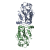
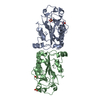
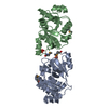
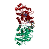
- PDB-1smn: IDENTIFICATION OF THE SERRATIA ENDONUCLEASE DIMER: STRUCTURAL BAS... -
+ Open data

- Basic information
| Entry | Database: PDB / ID: 1smn | ||||||
|---|---|---|---|---|---|---|---|
| Title | IDENTIFICATION OF THE SERRATIA ENDONUCLEASE DIMER: STRUCTURAL BASIS AND IMPLICATIONS FOR CATALYSIS | ||||||
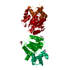
 Components Components | EXTRACELLULAR ENDONUCLEASE | ||||||
 Keywords Keywords |  ENDONUCLEASE / NUCLEASE / DNASE / RNASE / SUGAR-NONSPECIFIC NUCLEASE ENDONUCLEASE / NUCLEASE / DNASE / RNASE / SUGAR-NONSPECIFIC NUCLEASE | ||||||
| Function / homology |  Function and homology informationSerratia marcescens nuclease / endonuclease activity / nucleic acid binding / extracellular region / metal ion binding Function and homology informationSerratia marcescens nuclease / endonuclease activity / nucleic acid binding / extracellular region / metal ion bindingSimilarity search - Function | ||||||
| Biological species |  Serratia marcescens (bacteria) Serratia marcescens (bacteria) | ||||||
| Method | X-RAY DIFFRACTION / Resolution: 2.04 Å | ||||||
 Authors Authors | Miller, M.D. / Krause, K.L. | ||||||
 Citation Citation | Journal: Protein Sci. / Year: 1996 Title: Identification of the Serratia endonuclease dimer: structural basis and implications for catalysis. Authors: Miller, M.D. / Krause, K.L. #1: Journal: Nat.Struct.Biol. / Year: 1994Title: 2.1 Angstroms Structure of Serratia Endonuclease Suggests a Mechanism for Binding to Double-Stranded DNA Authors: Miller, M.D. / Tanner, J. / Alpaugh, M. / Benedik, M.J. / Krause, K.L. #2: Journal: J.Mol.Biol. / Year: 1991Title: Crystallization and Preliminary Crystallographic Analysis of a Novel Nuclease from Serratia Marcescens Authors: Miller, M.D. / Benedik, M.J. / Sullivan, M.J. / Shipley, M.C. / Krause, K.L. | ||||||
| History |
|
- Structure visualization
| Structure viewer | Molecule: MolmilJmol/JSmol |
|---|
- Downloads & links
-Download
| PDBx/mmCIF format | 1smn.cif.gz | 101.5 KB | Display | PDBx/mmCIF format |
|---|---|---|---|---|
| PDB format | pdb1smn.ent.gz | 83.2 KB | Display | PDB format |
| PDBx/mmJSON format | 1smn.json.gz | Tree view | PDBx/mmJSON format | |
| Others |  Other downloads Other downloads |
-Validation report
| Arichive directory | https://data.pdbj.org/pub/pdb/validation_reports/sm/1smnftp://data.pdbj.org/pub/pdb/validation_reports/sm/1smn | HTTPS FTP |
|---|
-Related structure data
| Similar structure data |
|---|


-Links
 PDBj
PDBj- Assembly
| Deposited unit | 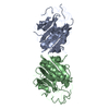
| ||||||||
|---|---|---|---|---|---|---|---|---|---|
| 1 |
| ||||||||
| Unit cell |
| ||||||||
| Noncrystallographic symmetry (NCS) | NCS oper: (Code: given Matrix: (-0.99989, -0.014803, -0.001089), Vector : Details | MTRIX THE TRANSFORMATIONS PRESENTED ON MTRIX RECORDS BELOW DESCRIBE NON-CRYSTALLOGRAPHIC RELATIONSHIPS AMONG THE VARIOUS DOMAINS IN THIS ENTRY. APPLYING THE APPROPRIATE MTRIX TRANSFORMATION TO THE RESIDUES LISTED FIRST WILL YIELD APPROXIMATE COORDINATES FOR THE RESIDUES LISTED SECOND. APPLIED TO TRANSFORMED TO MTRIX RESIDUES RESIDUES RMSD M1 A 5 .. A 245 B 5 .. B 245 0.131 | |
-Components
| #1: Protein | Mass: 26738.896 Da / Num. of mol.: 2 Source method: isolated from a genetically manipulated source Source: (gene. exp.) Serratia marcescens (bacteria) / Strain: SM6 / Gene: NUCA / Plasmid: PUC19NUC4OC / Gene (production host): NUCA / Production host: Serratia marcescens (bacteria) / References: UniProt: P13717, Serratia marcescens nuclease#2: Water | ChemComp-HOH / | Water Mass: 18.015 Da / Num. of mol.: 224 / Source method: isolated from a natural source / Formula: H2O Mass: 18.015 Da / Num. of mol.: 224 / Source method: isolated from a natural source / Formula: H2O |
|---|
-Experimental details
-Experiment
| Experiment | Method: X-RAY DIFFRACTION |
|---|
- Sample preparation
| Crystal | Density Matthews: 2.56 Å3/Da / Density % sol: 51.93 % Description: THIS STRUCTURE WAS SOLVED USING MULTIPLE ISOMORPHOUS REPLACEMENT WITH 5 DERIVATIVES. AFTER THE INITIAL MODEL WAS BUILT, THE DATA WAS REFINED AGAINST A NATIVE DATA SET COLLECTED FROM TWO CRYSTALS. | ||||||||||||||||||||||||||||||
|---|---|---|---|---|---|---|---|---|---|---|---|---|---|---|---|---|---|---|---|---|---|---|---|---|---|---|---|---|---|---|---|
| Crystal | *PLUS Density % sol: 52 % | ||||||||||||||||||||||||||||||
| Crystal grow | *PLUS pH: 8.2 / Method: microdialysisDetails: 2 can be substituted with 40 mM Tris-HCl, pH 8.2, 2 mM MgCl, 1 M NaCl2; or 100 mM NaPO4, pH 6.0. | ||||||||||||||||||||||||||||||
| Components of the solutions | *PLUS
|
-Data collection
| Diffraction |
| ||||||||||||
|---|---|---|---|---|---|---|---|---|---|---|---|---|---|
| Diffraction source |
| ||||||||||||
| Detector |
| ||||||||||||
| Radiation | Scattering type: x-ray | ||||||||||||
| Radiation wavelength | Wavelength: 1.54 Å / Relative weight: 1 | ||||||||||||
| Reflection | % possible obs: 93.7 % / Observed criterion σ(I): 0 |
- Processing
| Software |
| ||||||||||||||||||||||||||||||||||||||||||||||||||||||||||||||||||||||||||||||||
|---|---|---|---|---|---|---|---|---|---|---|---|---|---|---|---|---|---|---|---|---|---|---|---|---|---|---|---|---|---|---|---|---|---|---|---|---|---|---|---|---|---|---|---|---|---|---|---|---|---|---|---|---|---|---|---|---|---|---|---|---|---|---|---|---|---|---|---|---|---|---|---|---|---|---|---|---|---|---|---|---|---|
| Refinement | Resolution: 2.04→6 Å / σ(F): 2 Details: CRYST1 THERE IS STRONG PSEUDO-CENTERING ARISING FROM THE NON-CRYSTALLOGRAPHIC SYMMETRY. SHIFTING ONE MONOMER BY ONLY 1.4 ANGSTROMS WITH A 1 DEGREE ROTATION WOULD CHANGE THE SPACE GROUP TO I ...Details: CRYST1 THERE IS STRONG PSEUDO-CENTERING ARISING FROM THE NON-CRYSTALLOGRAPHIC SYMMETRY. SHIFTING ONE MONOMER BY ONLY 1.4 ANGSTROMS WITH A 1 DEGREE ROTATION WOULD CHANGE THE SPACE GROUP TO I 2 2 2. THIS STRUCTURE WAS SOLVED USING MULTIPLE ISOMORPHOUS REPLACEMENT WITH 5 DERIVATIVES. AFTER THE INITIAL MODEL WAS BUILT, THE DATA WAS REFINED AGAINST A NATIVE DATA SET COLLECTED FROM TWO CRYSTALS.
| ||||||||||||||||||||||||||||||||||||||||||||||||||||||||||||||||||||||||||||||||
| Displacement parameters | Biso mean: 11 Å2 | ||||||||||||||||||||||||||||||||||||||||||||||||||||||||||||||||||||||||||||||||
| Refine analyze | Luzzati coordinate error obs: 0.15 Å | ||||||||||||||||||||||||||||||||||||||||||||||||||||||||||||||||||||||||||||||||
| Refinement step | Cycle: LAST / Resolution: 2.04→6 Å
| ||||||||||||||||||||||||||||||||||||||||||||||||||||||||||||||||||||||||||||||||
| Refine LS restraints |
| ||||||||||||||||||||||||||||||||||||||||||||||||||||||||||||||||||||||||||||||||
| Refine LS restraints | *PLUS
|
