 Movie
Movie Controller

[English] 日本語
 Yorodumi
- PDB-1qae: THE ACTIVE SITE OF SERRATIA ENDONUCLEASE CONTAINS A CONSERVED MAG... -
+ Open data

- Basic information
| Entry | Database: PDB / ID: 1qae | ||||||
|---|---|---|---|---|---|---|---|
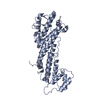
| Title | THE ACTIVE SITE OF SERRATIA ENDONUCLEASE CONTAINS A CONSERVED MAGNESIUM-WATER CLUSTER | ||||||
 Components Components | PROTEIN (EXTRACELLULAR ENDONUCLEASE) | ||||||
 Keywords Keywords | ENDONUCLEASE / NUCLEASE / DNASE / RNASE / SUGAR-NONSPECIFIC NUCLEASE / MAGNESIUM | ||||||
| Function / homology |  Function and homology information Function and homology informationSerratia marcescens nuclease / endonuclease activity / nucleic acid binding / hydrolase activity / extracellular region / metal ion binding Similarity search - Function | ||||||
| Biological species |  Serratia marcescens (bacteria) Serratia marcescens (bacteria) | ||||||
| Method |  X-RAY DIFFRACTION / Resolution: 2.05 Å X-RAY DIFFRACTION / Resolution: 2.05 Å | ||||||
 Authors Authors | Miller, M.D. / Krause, K.L. | ||||||
 Citation Citation | Journal: J.Mol.Biol. / Year: 1999 Title: The active site of Serratia endonuclease contains a conserved magnesium-water cluster. Authors: Miller, M.D. / Cai, J. / Krause, K.L. #1: Journal: Protein Sci. / Year: 1996Title: Identification of the Serratia Endonuclease Dimer: Structural Basis and Implications for Catalysis Authors: Miller, M.D. / Krause, K.L. #2: Journal: Nat.Struct.Biol. / Year: 1994Title: 2.1 Angstroms Structure of Serratia Endonuclease Suggests a Mechanism for Binding to Double-Stranded DNA Authors: Miller, M.D. / Tanner, J. / Alpaugh, M. / Benedik, M.J. / Krause, K.L. | ||||||
| History |
|
- Structure visualization
| Structure viewer | Molecule: MolmilJmol/JSmol |
|---|
- Downloads & links
-Download
| PDBx/mmCIF format | 1qae.cif.gz | 108.5 KB | Display | PDBx/mmCIF format |
|---|---|---|---|---|
| PDB format | pdb1qae.ent.gz | 83.6 KB | Display | PDB format |
| PDBx/mmJSON format | 1qae.json.gz | Tree view | PDBx/mmJSON format | |
| Others |  Other downloads Other downloads |
-Validation report
| Arichive directory | https://data.pdbj.org/pub/pdb/validation_reports/qa/1qaeftp://data.pdbj.org/pub/pdb/validation_reports/qa/1qae | HTTPS FTP |
|---|
-Related structure data
| Similar structure data |
|---|

-Links
 PDBj
PDBj- Assembly
| Deposited unit | 
| |||||||||||
|---|---|---|---|---|---|---|---|---|---|---|---|---|
| 1 |
| |||||||||||
| Unit cell |
| |||||||||||
| Noncrystallographic symmetry (NCS) | NCS domain:
NCS oper: (Code: given Matrix: (-0.999819, -0.018722, -0.003478), Vector: Details | MTRIX THE TRANSFORMATIONS PRESENTED ON MTRIX RECORDS BELOW DESCRIBE NON-CRYSTALLOGRAPHIC RELATIONSHIPS AMONG THE VARIOUS DOMAINS IN THIS ENTRY. APPLYING THE APPROPRIATE MTRIX TRANSFORMATION TO THE RESIDUES LISTED FIRST WILL YIELD APPROXIMATE COORDINATES FOR THE RESIDUES LISTED SECOND. APPLIED TO TRANSFORMED TO MTRIX RESIDUES RESIDUES RMSD M1 A 5 .. A 245 B 5 .. B 245 0.131 | |
-Components
| #1: Protein | Mass: 26738.896 Da / Num. of mol.: 2 Source method: isolated from a genetically manipulated source Source: (gene. exp.) Serratia marcescens (bacteria) / Strain: SM6 / Cellular location: EXTRACELLULARPlasmid details: PLASMID WHICH ENCODES THE GENE AND CARRIES AN OPERATOR CONSTITUTIVE MUTATION Plasmid: PUC19NUC4OC / Gene (production host): NUCA / Production host: Serratia marcescens (bacteria) / Strain (production host): PROTEASE DEFICIENT STRAIN / References: UniProt: P13717, Serratia marcescens nuclease#2: Chemical |   Mass: 24.305 Da / Num. of mol.: 2 / Source method: obtained synthetically / Formula: Mg Mass: 24.305 Da / Num. of mol.: 2 / Source method: obtained synthetically / Formula: Mg#3: Water | ChemComp-HOH / |  Mass: 18.015 Da / Num. of mol.: 251 / Source method: isolated from a natural source / Formula: H2O Mass: 18.015 Da / Num. of mol.: 251 / Source method: isolated from a natural source / Formula: H2OHas protein modification | Y | Sequence details | REFERENCE DATABASE: GENBANK ENTRY_NAME: SMANUCES | |
|---|
-Experimental details
-Experiment
| Experiment | Method: X-RAY DIFFRACTION / Number of used crystals: 1 |
|---|
- Sample preparation
| Crystal | Density Matthews: 2.57 Å3/Da / Density % sol: 52 % | ||||||||||||||||||||
|---|---|---|---|---|---|---|---|---|---|---|---|---|---|---|---|---|---|---|---|---|---|
| Crystal grow | Temperature: 277 K / Method: vapor diffusion, sitting drop / pH: 8.2 Details: CRYSTALLIZATION METHOD: VAPOR DIFFUSION SITTING DROP CRYSTALLIZATION TEMPERATURE: 277 K CRYSTALLIZATION PH: 4.6 CRYSTALS WERE GROWN FROM PEG 3350, AMMONIUM SULFATE, SODIUM ACETATE, AND ...Details: CRYSTALLIZATION METHOD: VAPOR DIFFUSION SITTING DROP CRYSTALLIZATION TEMPERATURE: 277 K CRYSTALLIZATION PH: 4.6 CRYSTALS WERE GROWN FROM PEG 3350, AMMONIUM SULFATE, SODIUM ACETATE, AND STORED IN A STABILIZATION SOLUTION CONTAINING 20% PEG 3000, 400mM GLYCINE, 40mM TRIS, PH8.2 (293 K), 20% PEG 3000, 10% PEG 400. THIS SOLUTION WAS EXCHANGED THREE TIMES TO MINIMIZE CARRY OVER FROM THE STORAGE SOLUTION. AFTER 3 DAYS IN THE MAGNESIUM STABILIZATION SOLUTION, THE CRYSTAL OF DIMENSION 0.4 X 0.4 X 0.3 MM**2 WAS MOUNTED IN A SILICONIZED SPECIAL GLASS CAPILLARY. , VAPOR DIFFUSION, SITTING DROP | ||||||||||||||||||||
| Crystal | *PLUS | ||||||||||||||||||||
| Crystal grow | *PLUS Temperature: 4 ℃ / pH: 4.6 / Method: other | ||||||||||||||||||||
| Components of the solutions | *PLUS
|
-Data collection
| Diffraction | Mean temperature: 288 K |
|---|---|
| Diffraction source | Source: ROTATING ANODE / Type: RIGAKU RU200 / Wavelength: 1.5418 |
| Detector | Type: ENRAF-NONIUS FAST / Detector: DIFFRACTOMETER / Date: Feb 2, 1998 |
| Radiation | Monochromator: GRAPHITE / Protocol: SINGLE WAVELENGTH / Monochromatic (M) / Laue (L): M / Scattering type: x-ray |
| Radiation wavelength | Wavelength: 1.5418 Å / Relative weight: 1 |
| Reflection | Resolution: 2.05→45 Å / Num. obs: 30574 / % possible obs: 84.3 % / Observed criterion σ(I): 0 / Redundancy: 3.1 % / Biso Wilson estimate: 6.6 Å2 / Rmerge(I) obs: 0.025 / Net I/σ(I): 40.87 |
| Reflection shell | Resolution: 2.05→2.1 Å / Redundancy: 1.6 % / Rmerge(I) obs: 0.045 / Mean I/σ(I) obs: 18.9 / % possible all: 20.4 |
| Reflection | *PLUS Num. measured all: 93785 |
- Processing
| Software |
| ||||||||||||||||||||||||||||||||||||||||||||||||||||||||||||||||||||||||||||||||
|---|---|---|---|---|---|---|---|---|---|---|---|---|---|---|---|---|---|---|---|---|---|---|---|---|---|---|---|---|---|---|---|---|---|---|---|---|---|---|---|---|---|---|---|---|---|---|---|---|---|---|---|---|---|---|---|---|---|---|---|---|---|---|---|---|---|---|---|---|---|---|---|---|---|---|---|---|---|---|---|---|---|
| Refinement | Starting model: 1.4 ANGSTROM STRUCTURE OF THE SERRATIAL ENDONUCLEASE (CAI, MILLER AND KRAUSE) WITH WATER AND ALTERNATE SIDE CHAIN CONFORMERS REMOVED AND THE SIDE CHAINS FOR THE ACTIVE SITE RESIDUES ...Starting model: 1.4 ANGSTROM STRUCTURE OF THE SERRATIAL ENDONUCLEASE (CAI, MILLER AND KRAUSE) WITH WATER AND ALTERNATE SIDE CHAIN CONFORMERS REMOVED AND THE SIDE CHAINS FOR THE ACTIVE SITE RESIDUES ARG 57, HIS 89, ASN 199 AND GLU 127 OMITTED Resolution: 2.05→50 Å / Rfactor Rfree error: 0.005 / Data cutoff high absF: 10000000 / Data cutoff low absF: 0 / Isotropic thermal model: RESTRAINED / Cross valid method: THROUGHOUT / σ(F): 2 Details: BULK SOLVENT MODEL USED, NCS B-FACTORS RESTRAINTS WERE APPLIED TO PROTEIN ATOMS AND PAIRS OF WATER MOLECULES WHICH DEVIATED LESS THAN 1.0 ANGSTROMS FROM NCS. THE PROTEIN ATOMS ARE IN GROUP 1 ...Details: BULK SOLVENT MODEL USED, NCS B-FACTORS RESTRAINTS WERE APPLIED TO PROTEIN ATOMS AND PAIRS OF WATER MOLECULES WHICH DEVIATED LESS THAN 1.0 ANGSTROMS FROM NCS. THE PROTEIN ATOMS ARE IN GROUP 1 AND THE WATER IS IN GROUP 2. WATER HOH 1 SITS ON THE NON-CRYSTALLOGRAPHIC 2-FOLD AXIS AND IT IS ITS OWN NCS MATE. THE WATER PAIRS IN GROUP 2 BEGIN WITH (HOH 2,HOH 3) AND END WITH (HOH 188, HOH 189). WATERS HOH 190 TO HOH 241 DO NOT HAVE NCS MATES WITHIN 1.0 ANGSTROMS. CRYST1 THERE IS STRONG PSEUDO-CENTERING ARISING FROM THE NON-CRYSTALLOGRAPHIC SYMMETRY. SHIFTING ONE MONOMER BY ONLY 1.4 ANGSTROMS WITH A 1 DEGREE ROTATION WOULD CHANGE THE SPACE GROUP TO I 2 2 2.
| ||||||||||||||||||||||||||||||||||||||||||||||||||||||||||||||||||||||||||||||||
| Displacement parameters | Biso mean: 14.8 Å2
| ||||||||||||||||||||||||||||||||||||||||||||||||||||||||||||||||||||||||||||||||
| Refine analyze |
| ||||||||||||||||||||||||||||||||||||||||||||||||||||||||||||||||||||||||||||||||
| Refinement step | Cycle: LAST / Resolution: 2.05→50 Å
| ||||||||||||||||||||||||||||||||||||||||||||||||||||||||||||||||||||||||||||||||
| Refine LS restraints |
| ||||||||||||||||||||||||||||||||||||||||||||||||||||||||||||||||||||||||||||||||
| Refine LS restraints NCS |
| ||||||||||||||||||||||||||||||||||||||||||||||||||||||||||||||||||||||||||||||||
| LS refinement shell | Resolution: 2.05→2.18 Å / Rfactor Rfree error: 0.019 / Total num. of bins used: 6
| ||||||||||||||||||||||||||||||||||||||||||||||||||||||||||||||||||||||||||||||||
| Xplor file |
| ||||||||||||||||||||||||||||||||||||||||||||||||||||||||||||||||||||||||||||||||
| Software | *PLUS Name: X-PLOR(ONLINE) / Version: 3.851 / Classification: refinement | ||||||||||||||||||||||||||||||||||||||||||||||||||||||||||||||||||||||||||||||||
| Refinement | *PLUS Rfactor obs: 0.167 / Rfactor Rfree: 0.21 | ||||||||||||||||||||||||||||||||||||||||||||||||||||||||||||||||||||||||||||||||
| Solvent computation | *PLUS | ||||||||||||||||||||||||||||||||||||||||||||||||||||||||||||||||||||||||||||||||
| Displacement parameters | *PLUS | ||||||||||||||||||||||||||||||||||||||||||||||||||||||||||||||||||||||||||||||||
| Refine LS restraints | *PLUS
|
