 Movie
Movie Controller

[English] 日本語
 Yorodumi
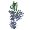
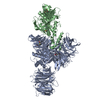
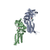
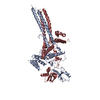
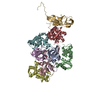
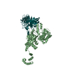
- PDB-1seb: COMPLEX OF THE HUMAN MHC CLASS II GLYCOPROTEIN HLA-DR1 AND THE BA... -
+ Open data

- Basic information
| Entry | Database: PDB / ID: 1seb | ||||||
|---|---|---|---|---|---|---|---|
| Title | COMPLEX OF THE HUMAN MHC CLASS II GLYCOPROTEIN HLA-DR1 AND THE BACTERIAL SUPERANTIGEN SEB | ||||||
 Components Components |
| ||||||
 Keywords Keywords | COMPLEX (MHC II/PEPTIDE/TOXIN) / HISTOCOMPATIBILITY ANTIGEN / MHC II / SUPERANTIGEN / ENTEROTOXIN PEPTIDE / TOXIN / COMPLEX (MHC II-PEPTIDE-TOXIN) COMPLEX | ||||||
| Function / homology |  Function and homology information Function and homology informationregulation of interleukin-4 production / regulation of interleukin-10 production / myeloid dendritic cell antigen processing and presentation / antigen processing and presentation of endogenous peptide antigen via MHC class II / positive regulation of T cell mediated immune response to tumor cell / autolysosome membrane / regulation of T-helper cell differentiation / positive regulation of CD4-positive, CD25-positive, alpha-beta regulatory T cell differentiation / MHC class II receptor activity / positive regulation of CD4-positive, alpha-beta T cell activation ...regulation of interleukin-4 production / regulation of interleukin-10 production / myeloid dendritic cell antigen processing and presentation / antigen processing and presentation of endogenous peptide antigen via MHC class II / positive regulation of T cell mediated immune response to tumor cell / autolysosome membrane / regulation of T-helper cell differentiation / positive regulation of CD4-positive, CD25-positive, alpha-beta regulatory T cell differentiation / MHC class II receptor activity / positive regulation of CD4-positive, alpha-beta T cell activation / antigen processing and presentation of peptide or polysaccharide antigen via MHC class II / positive regulation of memory T cell differentiation / positive regulation of monocyte differentiation / CD4 receptor binding / inflammatory response to antigenic stimulus / positive regulation of kinase activity / intermediate filament / transport vesicle membrane / polysaccharide binding / T-helper 1 type immune response / Translocation of ZAP-70 to Immunological synapse / Phosphorylation of CD3 and TCR zeta chains / positive regulation of insulin secretion involved in cellular response to glucose stimulus / humoral immune response / macrophage differentiation / negative regulation of type II interferon production / Generation of second messenger molecules / immunological synapse / PD-1 signaling / epidermis development / T cell receptor binding / negative regulation of T cell proliferation / detection of bacterium / negative regulation of inflammatory response to antigenic stimulus / MHC class II antigen presentation / trans-Golgi network membrane / clathrin-coated endocytic vesicle membrane / lumenal side of endoplasmic reticulum membrane / protein tetramerization / ER to Golgi transport vesicle membrane / structural constituent of cytoskeleton / cognition / peptide antigen assembly with MHC class II protein complex / MHC class II protein complex / positive regulation of T cell mediated cytotoxicity / peptide antigen binding / antigen processing and presentation of exogenous peptide antigen via MHC class II / endocytic vesicle membrane / Interferon gamma signaling / positive regulation of immune response / positive regulation of T cell activation / Downstream TCR signaling / MHC class II protein complex binding / early endosome membrane / late endosome membrane / T cell receptor signaling pathway / positive regulation of canonical NF-kappaB signal transduction / toxin activity / adaptive immune response / positive regulation of ERK1 and ERK2 cascade / positive regulation of MAPK cascade / positive regulation of viral entry into host cell / lysosome / positive regulation of protein phosphorylation / immune response / Golgi membrane / external side of plasma membrane / lysosomal membrane / positive regulation of DNA-templated transcription / cell surface / signal transduction / extracellular space / extracellular exosome / extracellular region / membrane / metal ion binding / plasma membrane Similarity search - Function | ||||||
| Biological species |  Homo sapiens (human) Homo sapiens (human)  Staphylococcus aureus (bacteria) Staphylococcus aureus (bacteria) | ||||||
| Method |  X-RAY DIFFRACTION / SYNCHROTRON / Resolution: 2.7 Å X-RAY DIFFRACTION / SYNCHROTRON / Resolution: 2.7 Å | ||||||
 Authors Authors | Jardetzky, T.S. / Brown, J.H. / Gorga, J.C. / Stern, L.J. / Urban, R.G. / Chi, Y.I. / Stauffacher, C. / Strominger, J.L. / Wiley, D.C. | ||||||
 Citation Citation | Journal: Nature / Year: 1994 Title: Three-dimensional structure of a human class II histocompatibility molecule complexed with superantigen. Authors: Jardetzky, T.S. / Brown, J.H. / Gorga, J.C. / Stern, L.J. / Urban, R.G. / Chi, Y.I. / Stauffacher, C. / Strominger, J.L. / Wiley, D.C. #1: Journal: Proc.Natl.Acad.Sci.USA / Year: 1996Title: Crystallographic Analysis of Endogenous Peptides Associated with Hla-Dr1 Suggests a Common, Polyproline II-Like Conformation for Bound Peptides Authors: Jardetzky, T.S. / Brown, J.H. / Gorga, J.C. / Stern, L.J. / Urban, R.G. / Strominger, J.L. / Wiley, D.C. | ||||||
| History |
|
- Structure visualization
| Structure viewer | Molecule: MolmilJmol/JSmol |
|---|
- Downloads & links
-Download
| PDBx/mmCIF format | 1seb.cif.gz | 233.7 KB | Display | PDBx/mmCIF format |
|---|---|---|---|---|
| PDB format | pdb1seb.ent.gz | 197.5 KB | Display | PDB format |
| PDBx/mmJSON format | 1seb.json.gz | Tree view | PDBx/mmJSON format | |
| Others |  Other downloads Other downloads |
-Validation report
| Summary document | 1seb_validation.pdf.gz | 410.2 KB | Display | wwPDB validaton report |
|---|---|---|---|---|
| Full document | 1seb_full_validation.pdf.gz | 448.5 KB | Display | |
| Data in XML | 1seb_validation.xml.gz | 27.9 KB | Display | |
| Data in CIF | 1seb_validation.cif.gz | 42.8 KB | Display | |
| Arichive directory | https://data.pdbj.org/pub/pdb/validation_reports/se/1sebftp://data.pdbj.org/pub/pdb/validation_reports/se/1seb | HTTPS FTP |
-Related structure data
| Similar structure data |
|---|
-Links
 PDBj
PDBj


- Assembly
| Deposited unit | 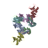
| ||||||||||||||||||||||||||||||||
|---|---|---|---|---|---|---|---|---|---|---|---|---|---|---|---|---|---|---|---|---|---|---|---|---|---|---|---|---|---|---|---|---|---|
| 1 |
| ||||||||||||||||||||||||||||||||
| Unit cell |
| ||||||||||||||||||||||||||||||||
| Noncrystallographic symmetry (NCS) | NCS oper:
| ||||||||||||||||||||||||||||||||
| Details | THE ASYMMETRIC UNIT CONTAINS A DIMER CONSISTING OF TWO HLA/DR-1 AND TWO SEB MOLECULES. THE DEPOSITORS PROVIDED A MONOMER AND THE TRANSFORMATIONS TO GENERATE THE OTHER MONOMER IN THE DIMER. THE PROTEIN DATA BANK GENERATED THE COMPLETE ASYMMETRIC UNIT USING THE TRANSFORMATIONS PROVIDED. CHAINS A, B, C, D WERE DEPOSITED AND CHAINS E, F, G, H WERE GENERATED USING THE TRANSFORMATIONS GIVEN ON MTRIX RECORDS BELOW. SEE REMARK 295 FOR ADDITIONAL DETAILS. |
-Components
| #1: Protein | Mass: 21084.826 Da / Num. of mol.: 2 / Fragment: EXTRACELLULAR DOMAIN Source method: isolated from a genetically manipulated source Source: (gene. exp.) Homo sapiens (human) / Cell line: LG-2 / Cell line (production host): LG-2 cells / Production host: Homo sapiens (human) / References: UniProt: P01903#2: Protein | Mass: 22324.938 Da / Num. of mol.: 2 / Fragment: EXTRACELLULAR DOMAIN Source method: isolated from a genetically manipulated source Source: (gene. exp.) Homo sapiens (human) / Cell line: LG-2 / Cell line (production host): LG-2 cells / Production host: Homo sapiens (human) / References: UniProt: P04229, UniProt: P01911*PLUS#3: Protein/peptide | Mass: 1124.378 Da / Num. of mol.: 2 / Source method: isolated from a natural source / Details: SUPERPOSITION OF MANY DIFFERENT PEPTIDES / Source: (natural) Homo sapiens (human)#4: Protein | Mass: 27793.309 Da / Num. of mol.: 2 / Source method: isolated from a natural source / Source: (natural) Staphylococcus aureus (bacteria) / Cell line: LG-2 / References: UniProt: P01552 |
|---|
-Experimental details
-Experiment
| Experiment | Method: X-RAY DIFFRACTION |
|---|
- Sample preparation
| Crystal | Density Matthews: 2.82 Å3/Da / Density % sol: 55 % | ||||||||||||||||||||||||||||||
|---|---|---|---|---|---|---|---|---|---|---|---|---|---|---|---|---|---|---|---|---|---|---|---|---|---|---|---|---|---|---|---|
| Crystal | *PLUS | ||||||||||||||||||||||||||||||
| Crystal grow | *PLUS Temperature: 25 ℃ / pH: 7.5 / Method: vapor diffusion | ||||||||||||||||||||||||||||||
| Components of the solutions | *PLUS
|
-Data collection
| Diffraction source | Source: SYNCHROTRON / Site: CHESS  / Beamline: F1 / Wavelength: 0.918 / Beamline: F1 / Wavelength: 0.918 |
|---|---|
| Detector | Type: KODAK / Detector: IMAGE PLATE / Date: Nov 1, 1991 |
| Radiation | Monochromatic (M) / Laue (L): M / Scattering type: x-ray |
| Radiation wavelength | Wavelength: 0.918 Å / Relative weight: 1 |
| Reflection | Num. obs: 40627 / % possible obs: 86 % / Redundancy: 3.3 % / Rmerge(I) obs: 0.057 |
| Reflection | *PLUS Highest resolution: 2.7 Å / Lowest resolution: 30 Å |
- Processing
| Software |
| ||||||||||||||||||||||||||||||||||||||||||||||||||||||||||||
|---|---|---|---|---|---|---|---|---|---|---|---|---|---|---|---|---|---|---|---|---|---|---|---|---|---|---|---|---|---|---|---|---|---|---|---|---|---|---|---|---|---|---|---|---|---|---|---|---|---|---|---|---|---|---|---|---|---|---|---|---|---|
| Refinement | Resolution: 2.7→6 Å / σ(F): 2 Details: THERE IS SOME EVIDENCE THAT THE SECOND SEB MOLECULE IS FOUND AT LOWER OCCUPANCY IN THE LATTICE. THE DR1:SEB STRUCTURE CONTAINS A BACKBONE MODEL (REFINED AS POLYALANINE) FOR ELECTRON DENSITY ...Details: THERE IS SOME EVIDENCE THAT THE SECOND SEB MOLECULE IS FOUND AT LOWER OCCUPANCY IN THE LATTICE. THE DR1:SEB STRUCTURE CONTAINS A BACKBONE MODEL (REFINED AS POLYALANINE) FOR ELECTRON DENSITY THAT CORRESPONDS TO A COMPLEX MIXTURE OF PEPTIDES THAT CO-PURIFY WITH THE HLA-DR1 MOLECULE (SEE REFERENCE 1 ABOVE FOR DETAILS). MHC CLASS II MOLECULES FORM VERY STABLE COMPLEXES WITH PEPTIDES AND, WHEN ISOLATED FROM HUMAN CELL LINES, HLA-DR1 MOLECULES HAVE BEEN SHOWN TO BE LOADED WITH A MIXTURE OF CELLULAR OR SERUM DERIVED PEPTIDES. THE NUMBER OF DIFFERENT PEPTIDES IS ESTIMATED TO BE ON THE ORDER OF THOUSANDS OF DIFFERENT SPECIES. IN THE CO-CRYSTAL OF HLA-DR1 WITH SEB, INTERPRETABLE ELECTRON DENSITY IS OBSERVED FOR THE BACKBONE OF A 13 AMINO ACID PEPTIDE WITH THE HLA-DR1 PEPTIDE-BINDING SITE. ALTHOUGH GOOD SIDE CHAIN DENSITY IS OBSERVED WITHIN THIS REGION, THE ELECTRON DENSITY COULD NOT BE UNAMBIGUOUSLY CORRELATED WITH ANY OF THE SEQUENCES DETERMINED FOR PEPTIDES ELUTED FROM PURIFIED HLA-DR1. THE ELECTRON DENSITY IS CONSISTENT WITH THE SUPERPOSITION OF MANY PEPTIDES WITH DIFFERENT SEQUENCES THAT ASSUME A COMMON CONFORMATION WITHIN THE HLA-DR1 PEPTIDE-BINDING SITE. THE POLYALANINE MODEL, THEREFORE, REPRESENTS A CONSENSUS CONFORMATION FOR PEPTIDE-BINDING TO HLA-DR1. IN THIS ENTRY THIS CHAIN IS PRESENTED AS CHAIN C (AND G) WITH THE RESIDUES NAMED "UNK".
| ||||||||||||||||||||||||||||||||||||||||||||||||||||||||||||
| Refinement step | Cycle: LAST / Resolution: 2.7→6 Å
| ||||||||||||||||||||||||||||||||||||||||||||||||||||||||||||
| Refine LS restraints |
|
