mature B cell differentiation involved in immune response / negative regulation of T cell differentiation in thymus / DNA recombinase complex / B cell homeostatic proliferation / endodeoxyribonuclease complex / pre-B cell allelic exclusion / negative regulation of T cell apoptotic process / positive regulation of organ growth / V(D)J recombination / negative regulation of thymocyte apoptotic process ...mature B cell differentiation involved in immune response / negative regulation of T cell differentiation in thymus / DNA recombinase complex / B cell homeostatic proliferation / endodeoxyribonuclease complex / pre-B cell allelic exclusion / negative regulation of T cell apoptotic process / positive regulation of organ growth / V(D)J recombination / negative regulation of thymocyte apoptotic process / phosphatidylinositol-3,4-bisphosphate binding / regulation of behavioral fear response / histone H3K4me3 reader activity / phosphatidylinositol-3,5-bisphosphate binding / regulation of T cell differentiation / positive regulation of T cell differentiation / T cell lineage commitment / B cell lineage commitment / T cell homeostasis / phosphatidylinositol-3,4,5-trisphosphate binding / T cell differentiation / protein autoubiquitination / phosphatidylinositol-4,5-bisphosphate binding / B cell differentiation / thymus development / phosphatidylinositol binding / visual learning / RING-type E3 ubiquitin transferase / ubiquitin-protein transferase activity / T cell differentiation in thymus / ubiquitin protein ligase activity / chromatin organization / endonuclease activity / DNA recombination / histone binding / sequence-specific DNA binding / Hydrolases; Acting on ester bonds / adaptive immune response / defense response to bacterium / hydrolase activity / chromatin binding / protein homodimerization activity / zinc ion binding / nucleoplasm / metal ion binding / identical protein binding / nucleus Similarity search - Function
Mass: 65.409 Da / Num. of mol.: 2 / Source method: obtained synthetically / Formula: Zn
-
Experimental details
-
Experiment
Experiment
Method: X-RAY DIFFRACTION / Number of used crystals: 1
-
Sample preparation
Crystal
Density Matthews: 3.46 Å3/Da / Density % sol: 64.48 %
Crystal grow
Temperature: 277 K / Method: vapor diffusion, hanging drop Details: 100 mM MES (pH 7.1), 10-15% PEG 3350, 200 mM tribasic ammonium citrate (pH 7.0), 100 mM KCl
In the structure databanks used in Yorodumi, some data are registered as the other names, "COVID-19 virus" and "2019-nCoV". Here are the details of the virus and the list of structure data.
Jan 31, 2019. EMDB accession codes are about to change! (news from PDBe EMDB page)
EMDB accession codes are about to change! (news from PDBe EMDB page)
The allocation of 4 digits for EMDB accession codes will soon come to an end. Whilst these codes will remain in use, new EMDB accession codes will include an additional digit and will expand incrementally as the available range of codes is exhausted. The current 4-digit format prefixed with “EMD-” (i.e. EMD-XXXX) will advance to a 5-digit format (i.e. EMD-XXXXX), and so on. It is currently estimated that the 4-digit codes will be depleted around Spring 2019, at which point the 5-digit format will come into force.
The EM Navigator/Yorodumi systems omit the EMD- prefix.
Related info.:Q: What is EMD? / ID/Accession-code notation in Yorodumi/EM Navigator
Yorodumi is a browser for structure data from EMDB, PDB, SASBDB, etc.
This page is also the successor to EM Navigator detail page, and also detail information page/front-end page for Omokage search.
The word "yorodu" (or yorozu) is an old Japanese word meaning "ten thousand". "mi" (miru) is to see.
Related info.:EMDB / PDB / SASBDB / Comparison of 3 databanks / Yorodumi Search / Aug 31, 2016. New EM Navigator & Yorodumi / Yorodumi Papers / Jmol/JSmol / Function and homology information / Changes in new EM Navigator and Yorodumi
 Movie
Movie Controller


 Open data
 Basic information
 Components
 Keywords
 Function and homology information
 X-RAY DIFFRACTION /
  Authors
 Citation
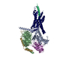
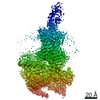
 Structure visualization
 Downloads & links
 Other downloads


 PDBj
PDBj


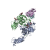
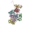
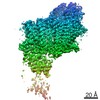
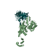
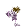
 Assembly
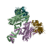
 Homo sapiens (human)
 Mass: 65.409 Da / Num. of mol.: 2 / Source method: obtained synthetically / Formula: Zn
 Sample preparation
 / Beamline: 22-ID / Wavelength: 1 Å
 Processing