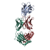
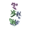
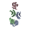
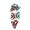
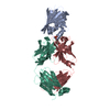
SEQUENCE CHAINS A AND B: NO SUITABLE SEQUENCE DATABASE REFERENCE WAS FOUND AT THE TIME OF ...SEQUENCE CHAINS A AND B: NO SUITABLE SEQUENCE DATABASE REFERENCE WAS FOUND AT THE TIME OF PROCESSING. CHAIN C: THE SEQUENCE LISTED IN THE SEQRES FOR CHAIN C IS THE PORTION OF THE FOLLOWING CONSTRUCT THAT WAS CLEARLY OBSERVED IN THE DENSITY: ANKLDSKKLTRSNGTTLEYSQITDADNATKAVETLKNSIKLEGSLVVGKTTVEIK EGTVTLKREIEKDGKVKVFLNDTAGSNKKTGKWEDSTSTLTISADSKKTKDLVFL TDGTITVQQYNTAGTSLEGSASEIKNLSELKNALK THE AUTHORS DO NOT KNOW IF THE PORTION OF THE CONSTRUCT THAT WAS NOT OBSERVED IN THE DENSITY WAS MISSING DUE TO PROTEOLYSIS, OR WAS DISORDERED IN THE CRYSTAL. CHAIN D: THE AUTHORS SAW AN EXTRA BETA STRAND OF DENSITY THAT WAS NOT CONTINUOUS WITH THE MAIN BODY OF DENSITY (I.E. THERE WAS NO DENSITY FOR A CONNECTING LOOP) AND THAT DID NOT SHOW CLEAR SIDECHAIN DENSITY. THIS STRAND PRESUMABLY ARISES FROM SOME PORTION OR PORTIONS OF THE REMAINING 'UNOBSERVED' SEQUENCE ABOVE, BUT SINCE THE REGISTER WAS UNKNOWN, THE AUTHORS MODELLED IT AS A POLYALANINE STRAND, LISTED IN THE SEQRES AND COORDINATES AS UNK, UNKNOWN RESIDUE.
In the structure databanks used in Yorodumi, some data are registered as the other names, "COVID-19 virus" and "2019-nCoV". Here are the details of the virus and the list of structure data.
Jan 31, 2019. EMDB accession codes are about to change! (news from PDBe EMDB page)
EMDB accession codes are about to change! (news from PDBe EMDB page)
The allocation of 4 digits for EMDB accession codes will soon come to an end. Whilst these codes will remain in use, new EMDB accession codes will include an additional digit and will expand incrementally as the available range of codes is exhausted. The current 4-digit format prefixed with “EMD-” (i.e. EMD-XXXX) will advance to a 5-digit format (i.e. EMD-XXXXX), and so on. It is currently estimated that the 4-digit codes will be depleted around Spring 2019, at which point the 5-digit format will come into force.
The EM Navigator/Yorodumi systems omit the EMD- prefix.
Related info.:Q: What is EMD? / ID/Accession-code notation in Yorodumi/EM Navigator
Yorodumi is a browser for structure data from EMDB, PDB, SASBDB, etc.
This page is also the successor to EM Navigator detail page, and also detail information page/front-end page for Omokage search.
The word "yorodu" (or yorozu) is an old Japanese word meaning "ten thousand". "mi" (miru) is to see.
Related info.:EMDB / PDB / SASBDB / Comparison of 3 databanks / Yorodumi Search / Aug 31, 2016. New EM Navigator & Yorodumi / Yorodumi Papers / Jmol/JSmol / Function and homology information / Changes in new EM Navigator and Yorodumi
 Movie
Movie Controller

 Yorodumi
 Open data
 Basic information
 Components
 Keywords
 Function and homology information
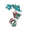
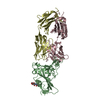
 Borrelia burgdorferi (Lyme disease spirochete)
 X-RAY DIFFRACTION /
  Authors
 Citation
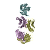
 Structure visualization
 Downloads & links
 Other downloads


 PDBj
PDBj


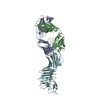
 Assembly
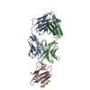
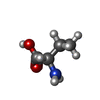
 Mass: 18.015 Da / Num. of mol.: 144 / Source method: isolated from a natural source / Formula: H2O
 Sample preparation
 / Beamline: X12C / Wavelength: 0.98 Å
 Processing